EMD-14533

Single-particle
3.5 Å
EMD-14533 Deposition: 15/03/2022
Map released: 29/06/2022
Last modified: 15/03/2023
Overview 3D View Sample Experiment Validation Additional data Links

EMD-14533

Archive Files (Depositor)

Primary 3D volume (map.gz) Half-map 1 Half-map 2 (map.gz) Experimental metadata (xml) Experimental metadata (cif.gz)

Validation (wwPDB)

Map-only validation report (pdf.gz) Map-7z7i summary report (pdf.gz) Map-7z7i FULL report (pdf.gz)

EMDB Annotations

EMICSS entry mapping (xml)

EMDB Files

VA raw map (map) VA resolution mask (mrc)

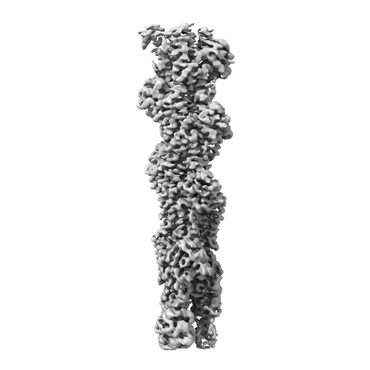
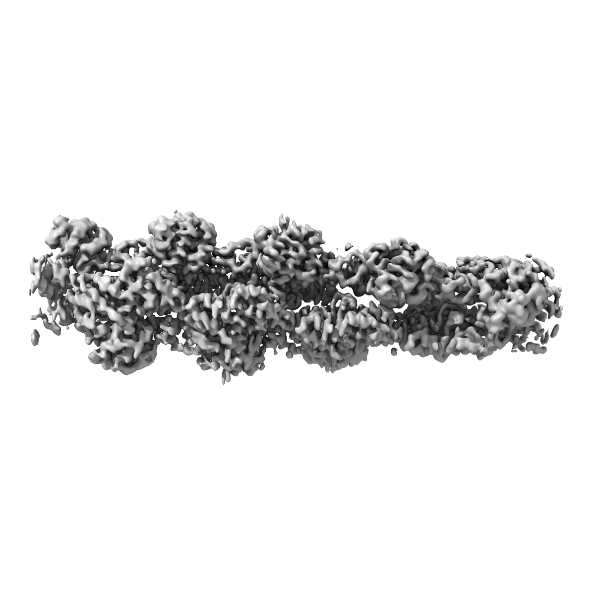
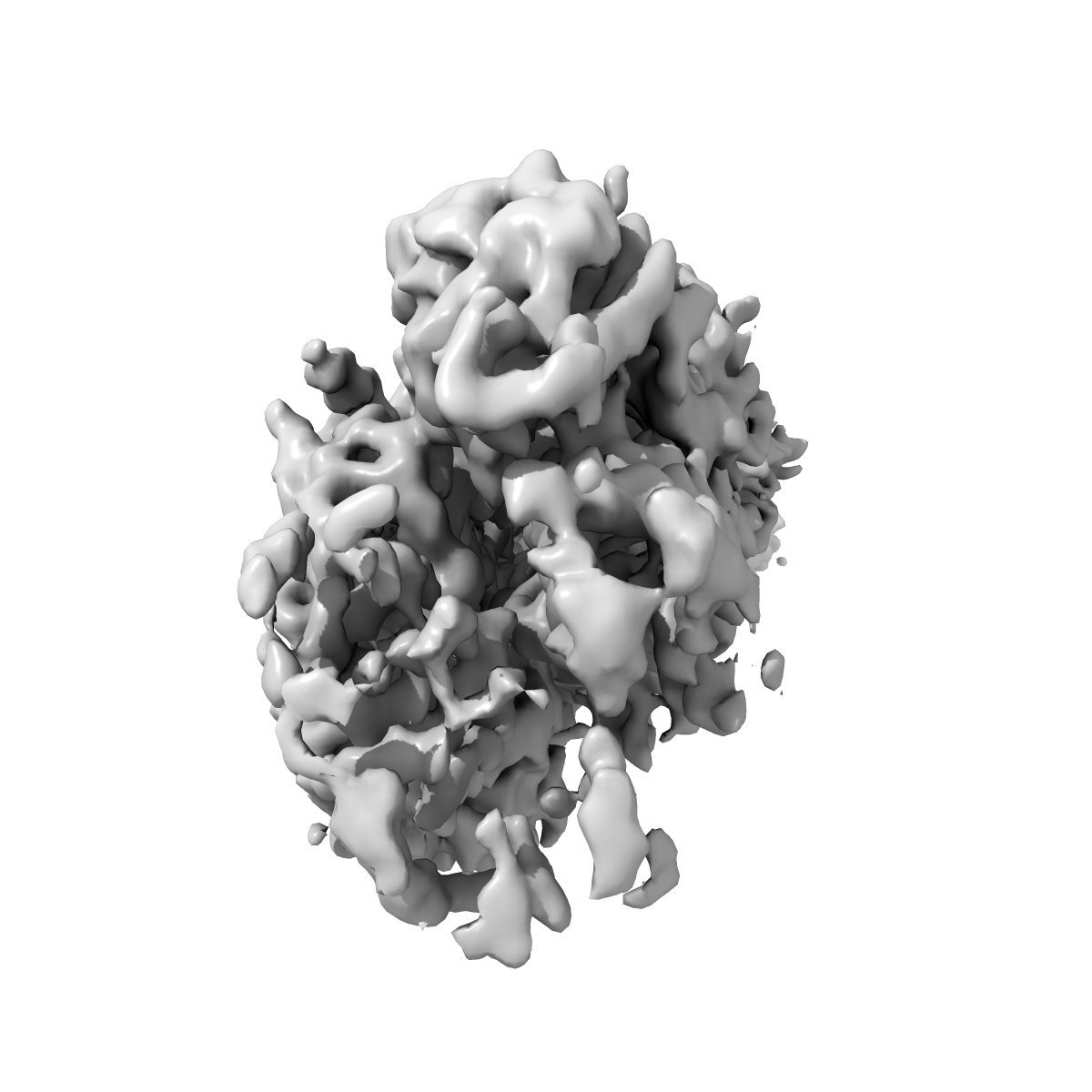
Structure of ADP-ribosylated F-actin

EMD-14533

Single-particle
3.5 Å
EMD-14533 Deposition: 15/03/2022
Map released: 29/06/2022
Last modified: 15/03/2023
Overview 3D View Sample Experiment Validation Additional data Links
Sample Organism: rabbit
Sample: Structure of ADP-ribosylated F-actin
Fitted models: 7z7i

Deposition Authors: Belyy A , Raunser S
Mechanism of threonine ADP-ribosylation of F-actin by a Tc toxin.
Belyy A , Lindemann F , Roderer D , Funk J, Bardiaux B , Protze J , Bieling P , Oschkinat H , Raunser S
(2022) Nat Commun , 13 , 4202 - 4202
PUBMED: 35858890
DOI: doi:10.1038/s41467-022-31836-w
ISSN: 2041-1723
Grant Support: