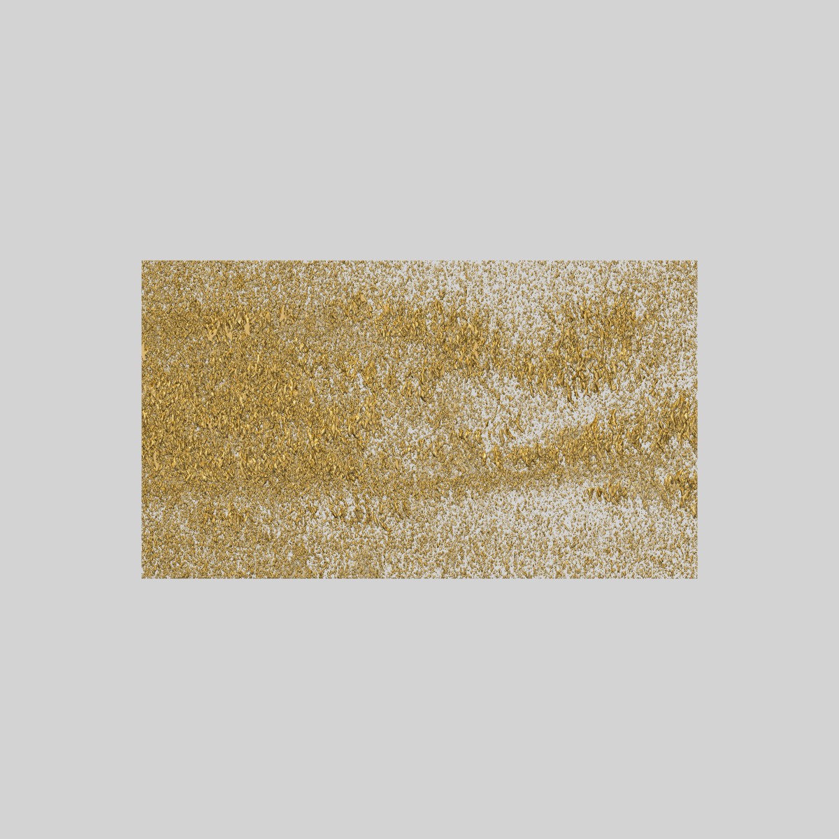
EMD-7880
Infectious microvesicles (Class 1 and 3) from poliovirus-infected HeLa cells, displaying clusters of virions and additional inner vesicular structures
EMD-7880
Tomography Deposition: 15/05/2018
Map released: 15/05/2019
Last modified: 27/05/2020
Concentration: 1.5
mg/mL
Buffer
pH: 7.0
Details: 1% glutaraldehyde in 1x PBS
Details: 1% glutaraldehyde in 1x PBS
Grid
Vitrification
Cryogen name: ETHANE
Chamber humidity: 100%
Chamber temperature: 283.15 K
Instrument: FEI VITROBOT MARK II
Chamber humidity: 100%
Chamber temperature: 283.15 K
Instrument: FEI VITROBOT MARK II
Fiducial Markers
| Manufacturer | Fiducial type | Diameter |
|---|---|---|
| Ted Pella Inc. | - | 10 nanometer |
Sectioning
Details: NO SECTIONING
Microscope: JEOL 2200FS
Illumination mode: FLOOD BEAM
Imaging mode: BRIGHT FIELD
Electron source: FIELD EMISSION GUN
Acceleration voltage: 200 kV
Illumination mode: FLOOD BEAM
Imaging mode: BRIGHT FIELD
Electron source: FIELD EMISSION GUN
Acceleration voltage: 200 kV
Image Recording
[1]
Final
reconstruction
Number of images used:
62
Software
[1]
| Name | Version | Details |
|---|---|---|
| ETomo | - | - |
Format: CCP4
Data type: IMAGE STORED AS SIGNED BYTE
Annotation details: Infectious microvesicles secreted from poliovirus-infected HeLa cells, displaying clusters of virions and additional inner vesicular structures
Data type: IMAGE STORED AS SIGNED BYTE
Annotation details: Infectious microvesicles secreted from poliovirus-infected HeLa cells, displaying clusters of virions and additional inner vesicular structures
⬡ Geometry
| X | Y | Z | |
|---|---|---|---|
| Origin | -886 | -2414 | 442 |
| Dimensions (px) | 753 | 843 | 431 |
| Dimensions (Å) | 3019.5303 | 3380.4302 | 1728.31 |
| Voxel size (Å) | 4.01 | 4.01 | 4.01 |
Contour list
| Primary | Level | Source |
|---|---|---|
| True | - | AUTHOR |