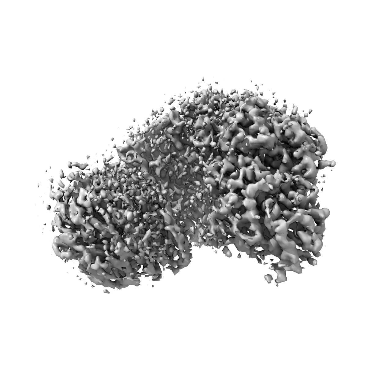
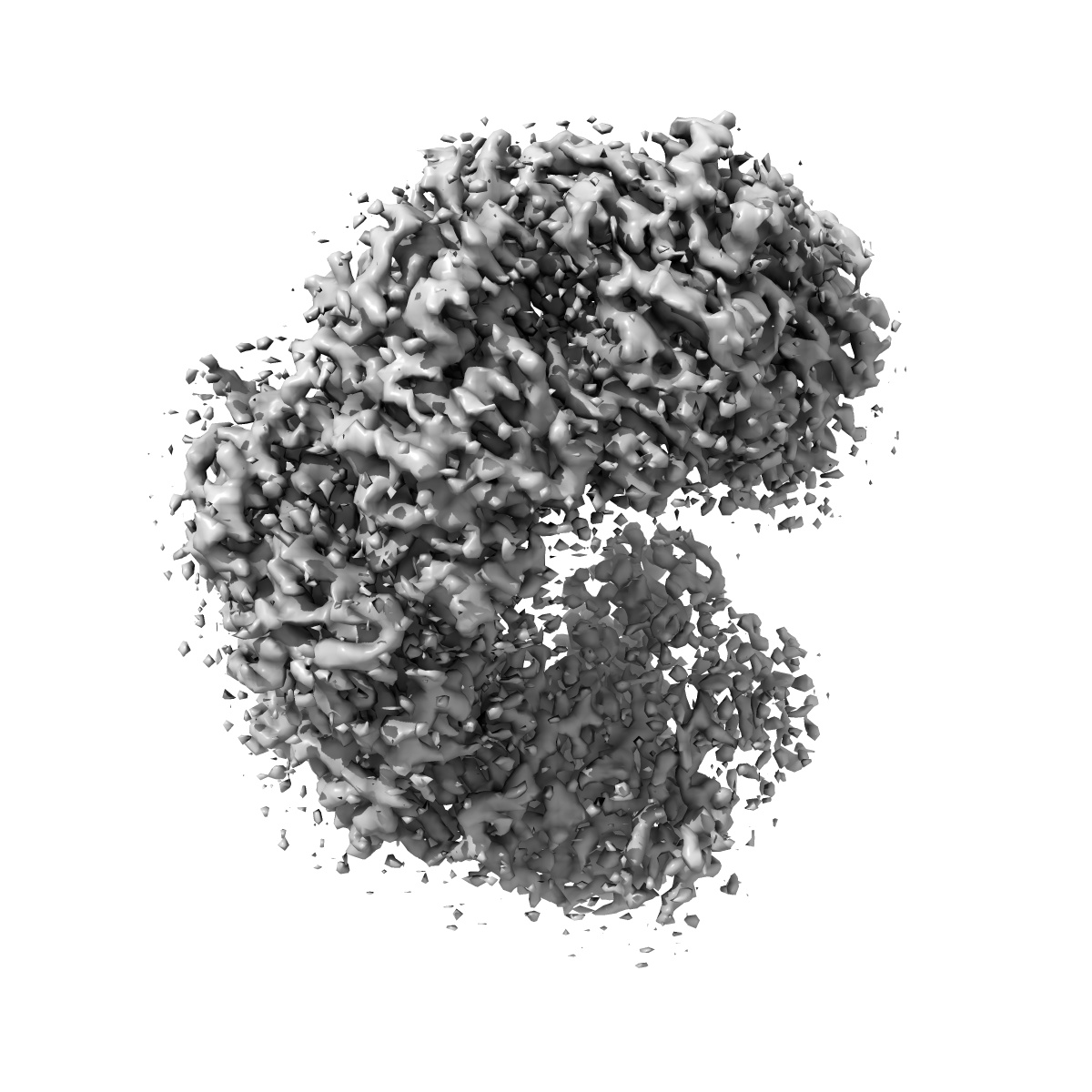
EMD-6729
Anti-CRISPR proteins AcrF1/2 bound to Csy surveillance complex with a 32nt spacer crRNA backbone region
EMD-6729
Single-particle3.8 Å
 Deposition: 11/05/2017
Map released: 10/01/2018
Last modified: 27/03/2024
Concentration: 0.4
mg/mL
Buffer
pH: 7.5
Grid
Vitrification
Cryogen name: ETHANE
Chamber humidity: 100%
Chamber temperature: 277 K
Instrument: FEI VITROBOT MARK IV
Chamber humidity: 100%
Chamber temperature: 277 K
Instrument: FEI VITROBOT MARK IV
Microscope: FEI TITAN KRIOS
Illumination mode: FLOOD BEAM
Imaging mode: BRIGHT FIELD
Electron source: FIELD EMISSION GUN
Acceleration voltage: 300 kV
Illumination mode: FLOOD BEAM
Imaging mode: BRIGHT FIELD
Electron source: FIELD EMISSION GUN
Acceleration voltage: 300 kV
Image Recording
[1]
Detector model:
GATAN K2 SUMMIT (4k x 4k)
Detector mode: SUPER-RESOLUTION
Average exposure time: 0.2 s
Average electron dose per image: 1.0 e/Å2
Detector mode: SUPER-RESOLUTION
Average exposure time: 0.2 s
Average electron dose per image: 1.0 e/Å2
Final
reconstruction
Resolution: 3.8
Å
(
BY AUTHOR)
Resolution method: FSC 0.143 CUT-OFF
Number of images used: 155099
Resolution method: FSC 0.143 CUT-OFF
Number of images used: 155099
⌯ Applied Symmetry
Point group:
C1
Software
[1]
| Name | Version | Details |
|---|---|---|
| RELION | 2.0 | - |
Startup model
[1]
⦨ Initial angle
assignment
⦩ Final angle assignment
Format: CCP4
Data type: IMAGE STORED AS FLOATING POINT NUMBER (4 BYTES)
Data type: IMAGE STORED AS FLOATING POINT NUMBER (4 BYTES)
⬡ Geometry
| X | Y | Z | |
|---|---|---|---|
| Origin | 0 | 0 | 0 |
| Dimensions (px) | 180 | 180 | 180 |
| Dimensions (Å) | 233.99998 | 233.99998 | 233.99998 |
| Voxel size (Å) | 1.3 | 1.3 | 1.3 |
Contour list
| Primary | Level | Source |
|---|---|---|
| True | 0.033 | AUTHOR |