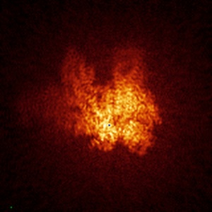
EMD-6479
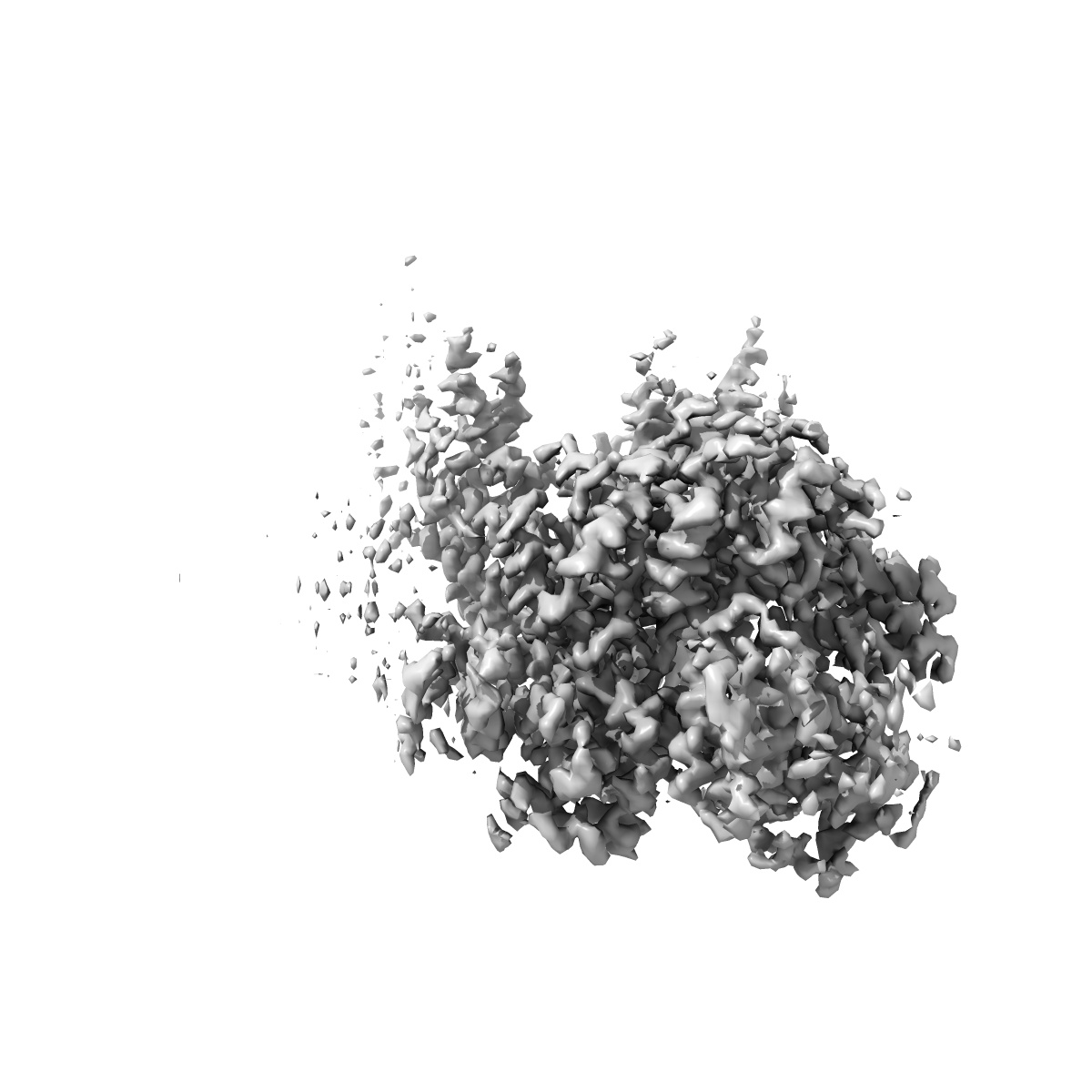
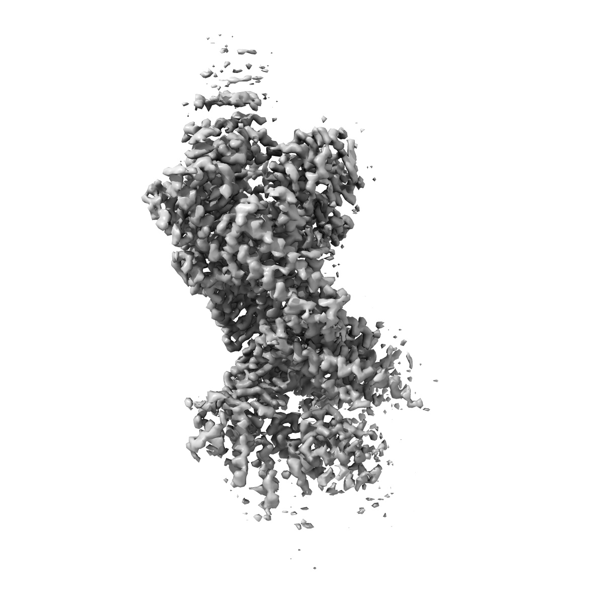
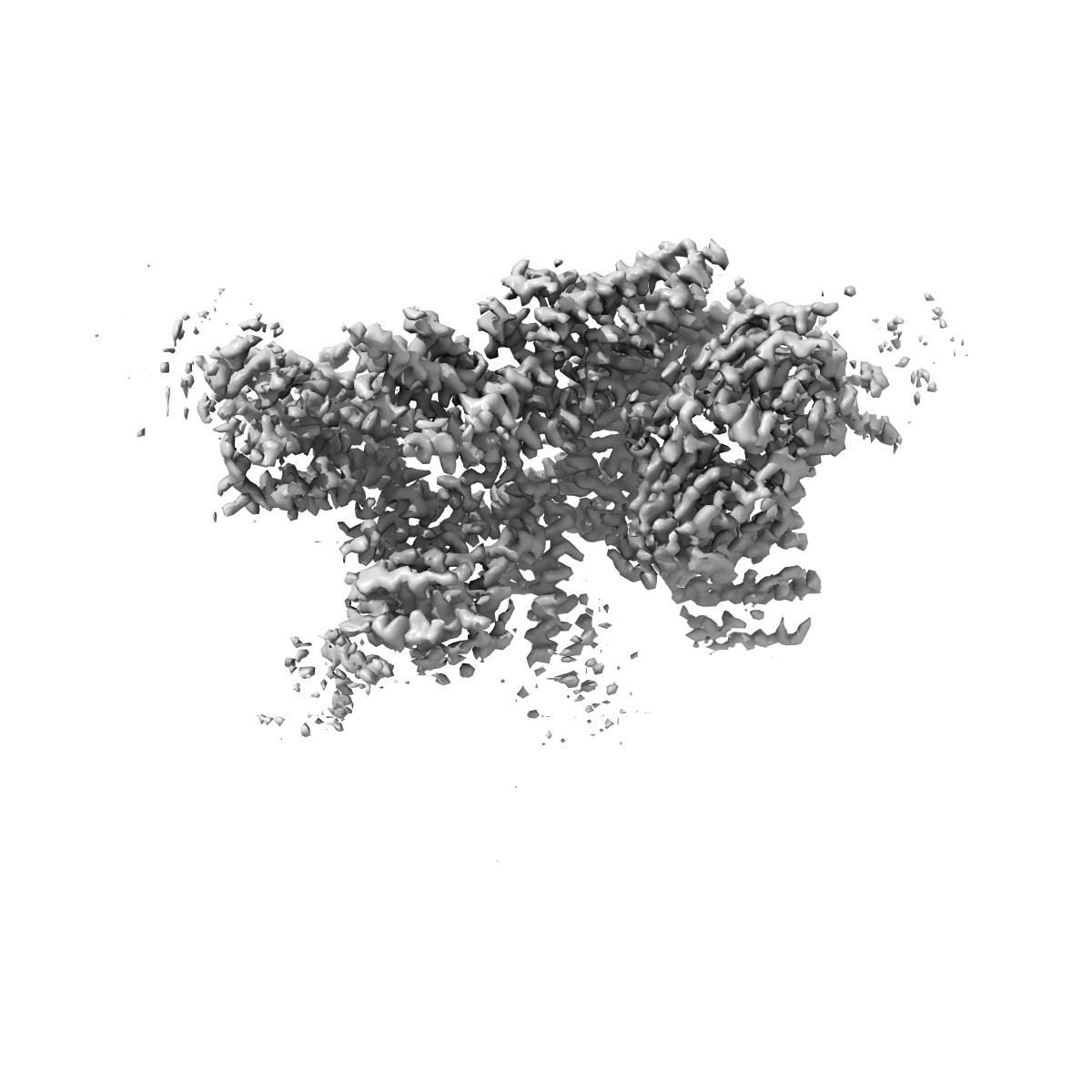
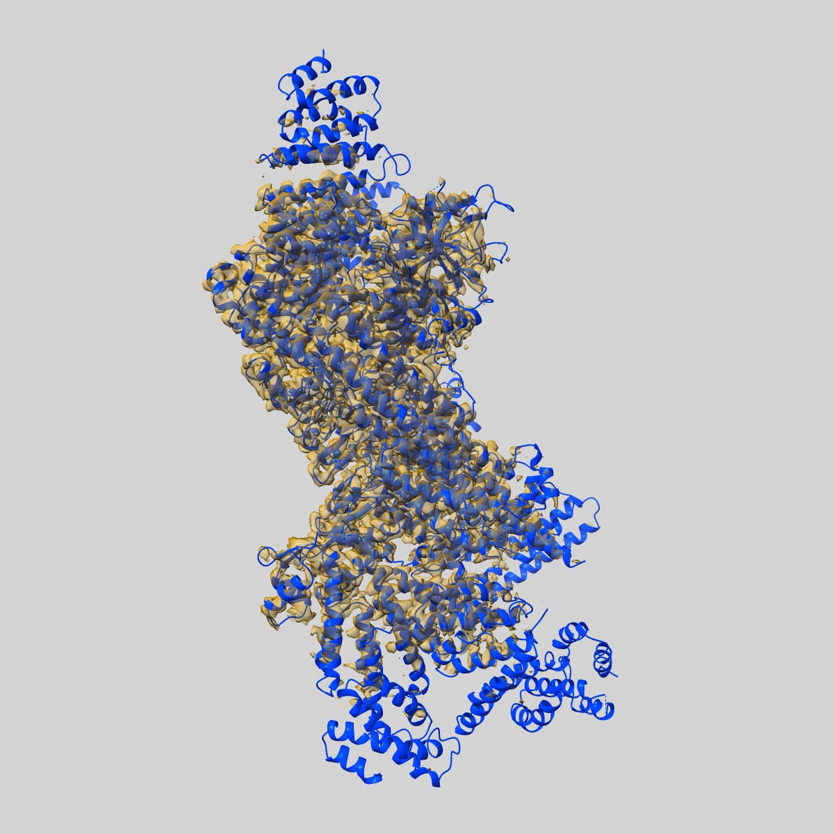
Structure of the yeast 26S proteasome lid sub-complex
EMD-6479
Single-particle3.5 Å
 Deposition: 11/10/2015
Map released: 20/01/2016
Last modified: 11/05/2016
Concentration: 2.5
mg/mL
Buffer
pH: 7.5
Details: 50 mM HEPES, 100 mM NaCl, 100 mM KCl, 1 mM TCEP
Details: 50 mM HEPES, 100 mM NaCl, 100 mM KCl, 1 mM TCEP
Grid
Details: Sample was applied directly to plasma-cleaned holey carbon C-flat grids (400 mesh, 1.2 micrometer holes).
Vitrification
Cryogen name: ETHANE
Chamber humidity: 88%
Chamber temperature: 85 K
Instrument: HOMEMADE PLUNGER
Method: 4 microliters of sample was applied to the grid, blotted for 2 seconds, and plunged into liquid ethane.
Details: Manual plunging was performed in a cold room.
Chamber humidity: 88%
Chamber temperature: 85 K
Instrument: HOMEMADE PLUNGER
Method: 4 microliters of sample was applied to the grid, blotted for 2 seconds, and plunged into liquid ethane.
Details: Manual plunging was performed in a cold room.
Microscope: FEI TITAN KRIOS
Illumination mode: FLOOD BEAM
Imaging mode: BRIGHT FIELD
Electron source: FIELD EMISSION GUN
Acceleration voltage: 300 kV
Nominal CS: 2.7 mm
Nominal defocus: 1.6 µm - 3.2 µm
Nominal magnification: 22500.0
Calibrated magnification: 38168.0
Specimen holder model: FEI TITAN KRIOS AUTOGRID HOLDER
Alignment procedure: LEGACY (Astigmatism: Objective lens astigmatism was corrected at a nominal magnification of 22,500., Electron beam tilt params: )
Details: Micrographs were collected in super-resolution mode with a total frame count of 38 and total exposure time of 7.6 seconds.
Temperature
Minimum: 85
K
Average: 87.5 K
Maximum: 90 K
Average: 87.5 K
Maximum: 90 K
Image Recording
[1]
Detector category:
CCD
Detector model: GATAN K2 (4k x 4k)
Number of real images: 3432
Average electron dose per image: 43.8 e/Å2
Details: Micrographs were collected as movies using super-resolution mode with the Gatan K2 Summit direct electron detector
Detector model: GATAN K2 (4k x 4k)
Number of real images: 3432
Average electron dose per image: 43.8 e/Å2
Details: Micrographs were collected as movies using super-resolution mode with the Gatan K2 Summit direct electron detector
Details: Image pre-processing was performed using Appion. 3D classification and reconstruction was performed with RELION.
Final
reconstruction
Resolution: 3.5
Å
(
BY AUTHOR)
Resolution method: OTHER
Number of images used: 109396
Algorithm: OTHER
Details: 3D classification was performed to identify the best 109,396 particles from an initial data set of 254,112.
Resolution method: OTHER
Number of images used: 109396
Algorithm: OTHER
Details: 3D classification was performed to identify the best 109,396 particles from an initial data set of 254,112.
⌯ Applied Symmetry
Point group:
C1
Software
[1]
| Name | Version | Details |
|---|---|---|
| Appion, CTFFIND3, FindEM, RELION | - | - |
CTF correction
Details: Whole micrograph
Format: CCP4
Data type: IMAGE STORED AS FLOATING POINT NUMBER (4 BYTES)
Annotation details: Reconstruction of the yeast proteasome lid sub-complex
Details: ::::EMDATABANK.org::::EMD-6479::::
Data type: IMAGE STORED AS FLOATING POINT NUMBER (4 BYTES)
Annotation details: Reconstruction of the yeast proteasome lid sub-complex
Details: ::::EMDATABANK.org::::EMD-6479::::
⬡ Geometry
| X | Y | Z | |
|---|---|---|---|
| Origin | 0 | 0 | 0 |
| Dimensions (px) | 160 | 160 | 160 |
| Dimensions (Å) | 209.59999 | 209.59999 | 209.59999 |
| Voxel size (Å) | 1.31 | 1.31 | 1.31 |
Contour list
| Primary | Level | Source |
|---|---|---|
| True | 0.0642 | AUTHOR |