EMD-6337
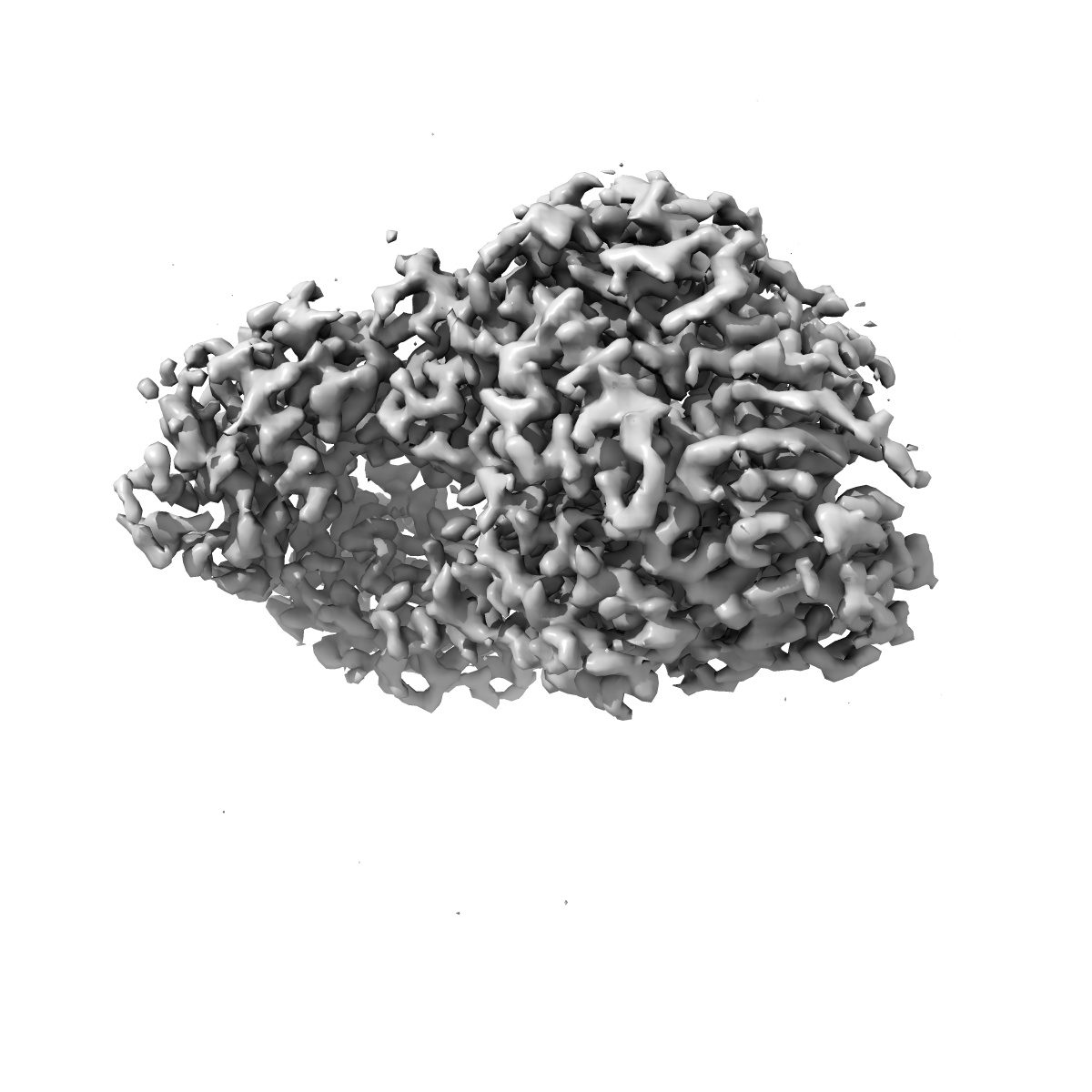
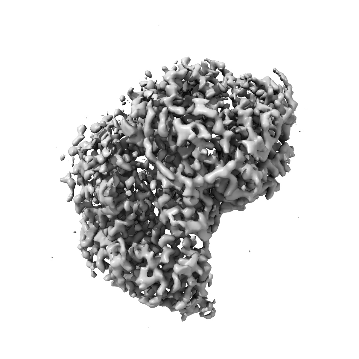
Structure of the L-protein of vesicular stomatitis virus from electron cryomicroscopy
EMD-6337
Single-particle3.8 Å
 Deposition: 07/05/2015
Map released: 20/05/2015
Last modified: 19/08/2015
Concentration: 0.35
mg/mL
Buffer
pH: 7.4
Details: 25 mM HEPES, 250 mM NaCl, 6 mM MgSO4, 0.5 mM TCEP
Details: 25 mM HEPES, 250 mM NaCl, 6 mM MgSO4, 0.5 mM TCEP
Grid
Details: 400 mesh Quantifoil R1.2/1.3 Cu grid
Vitrification
Cryogen name: ETHANE
Chamber humidity: 65%
Instrument: FEI VITROBOT MARK I
Method: Blot time 2 seconds, drain time 1 second before plunging
Chamber humidity: 65%
Instrument: FEI VITROBOT MARK I
Method: Blot time 2 seconds, drain time 1 second before plunging
Microscope: FEI TECNAI F20
Illumination mode: FLOOD BEAM
Imaging mode: BRIGHT FIELD
Electron source: FIELD EMISSION GUN
Acceleration voltage: 200 kV
Nominal CS: 2.0 mm
Nominal defocus: 0.9 µm - 2.3 µm
Nominal magnification: 29000.0
Calibrated magnification: 40410.0
Specimen holder model: GATAN LIQUID NITROGEN
Specimen holder details: CT3500
Details: Beam intensity: 8 electrons/pixel/s Movie mode: 30 frames, 5 frames/s
Image Recording
[1]
Detector category:
CCD
Detector model: GATAN K2 (4k x 4k)
Sampling interval: 5 µm
Number of real images: 1272
Average electron dose per image: 31 e/Å2
Details: Images are the sums of all 30 aligned movie frames (high dose) or frames 3 - 12 (low-dose).
Detector model: GATAN K2 (4k x 4k)
Sampling interval: 5 µm
Number of real images: 1272
Average electron dose per image: 31 e/Å2
Details: Images are the sums of all 30 aligned movie frames (high dose) or frames 3 - 12 (low-dose).
Details: An initial map was obtained with EMAN2, IMAGIC, and TIGRIS. CTF was determined using CTFFIND3. Refinement and classification were done using Frealign.
Final
reconstruction
Resolution: 3.8
Å
(
BY AUTHOR)
Resolution method: OTHER
Number of images used: 74940
Algorithm: OTHER
Details: The final map represents the best class out of three classes.
Resolution method: OTHER
Number of images used: 74940
Algorithm: OTHER
Details: The final map represents the best class out of three classes.
⌯ Applied Symmetry
Point group:
C1
Software
[1]
| Name | Version | Details |
|---|---|---|
| EMAN2, IMAGIC, Frealign | - | - |
Final 2D classification
Number of classes:
3
CTF correction
Details: Each particle
Format: CCP4
Data type: IMAGE STORED AS FLOATING POINT NUMBER (4 BYTES)
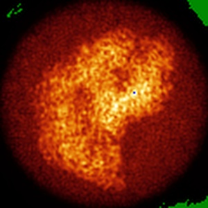
Annotation details: Reconstruction of the L-protein of vesicular stomatitis virus
Details: ::::EMDATABANK.org::::EMD-6337::::
Data type: IMAGE STORED AS FLOATING POINT NUMBER (4 BYTES)
Annotation details: Reconstruction of the L-protein of vesicular stomatitis virus
Details: ::::EMDATABANK.org::::EMD-6337::::
⬡ Geometry
| X | Y | Z | |
|---|---|---|---|
| Origin | 0 | 0 | 0 |
| Dimensions (px) | 120 | 120 | 120 |
| Dimensions (Å) | 148.44 | 148.44 | 148.44 |
| Voxel size (Å) | 1.237 | 1.237 | 1.237 |
Contour list
| Primary | Level | Source |
|---|---|---|
| True | 1.2 | AUTHOR |