EMD-5367

Helical reconstruction
15.0 Å
EMD-5367 Deposition: 09/12/2011
Map released: 26/03/2012
Last modified: 03/04/2012
Overview 3D View Sample Experiment Validation Additional data Links

EMD-5367

Archive Files (Depositor)

Primary 3D volume (map.gz) Experimental metadata (xml) Experimental metadata (cif.gz)

Validation (wwPDB)

Map-only validation report (pdf.gz)

EMDB Annotations

EMICSS entry mapping (xml)

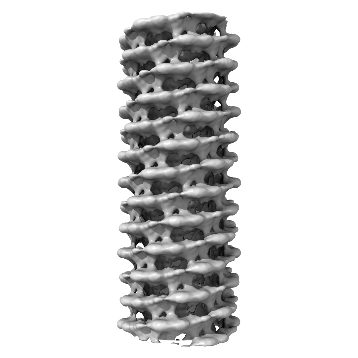
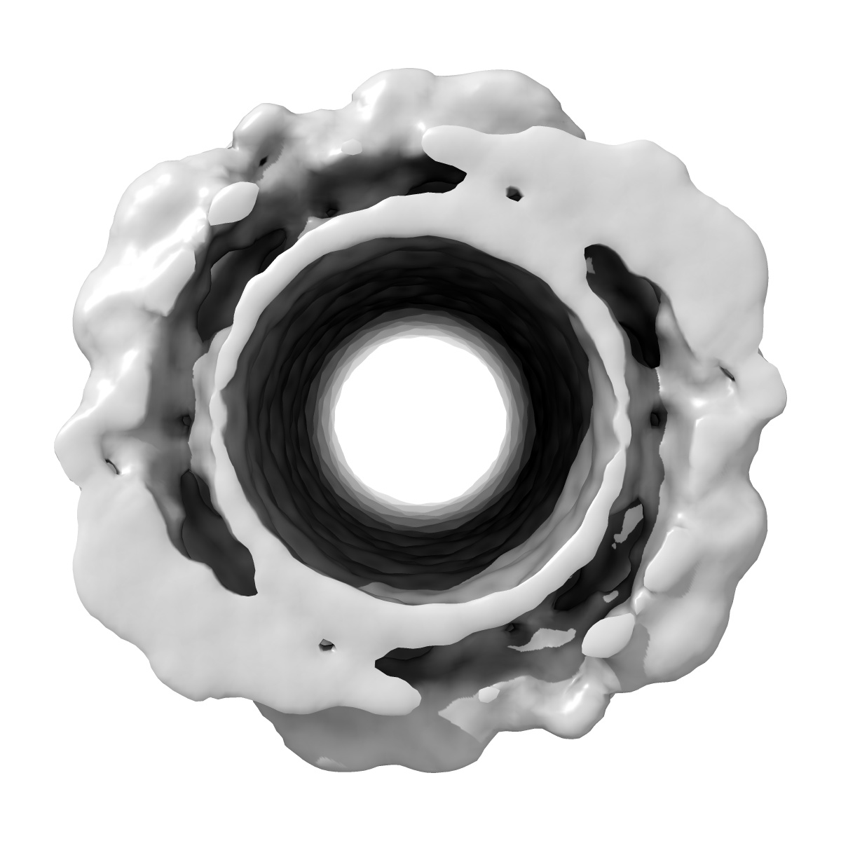
Structure of endophilin bound to a membrane tubule with a diameter of 28nm

EMD-5367

Helical reconstruction
15.0 Å
EMD-5367 Deposition: 09/12/2011
Map released: 26/03/2012
Last modified: 03/04/2012
Overview 3D View Sample Experiment Validation Additional data Links
Sample Organism: Rattus norvegicus
Sample: Structure of Endophilin bound to the bilayer

Deposition Authors: Mim C, Cui H, Gawronski-Salerno JA, Frost A, Lyman E, Voth GA, Unger VM
Structural basis of membrane bending by the N-BAR protein endophilin.
Mim C , Cui H, Gawronski-Salerno JA, Frost A , Lyman E, Voth GA, Unger VM
(2012) Cell , 149 , 137 - 145