EMD-3531
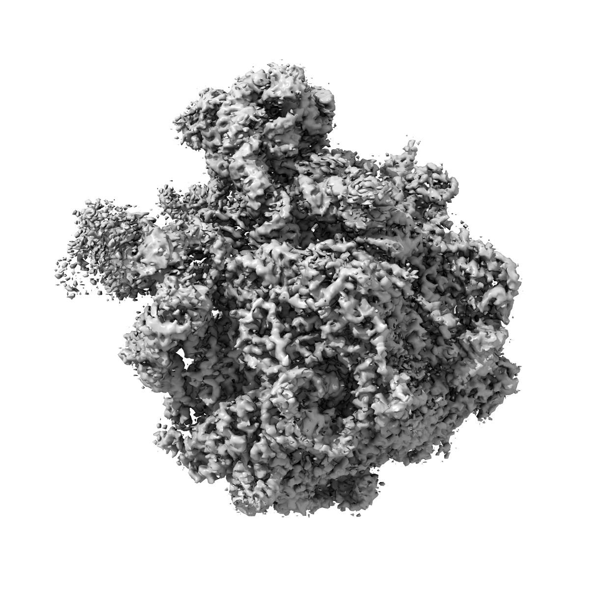
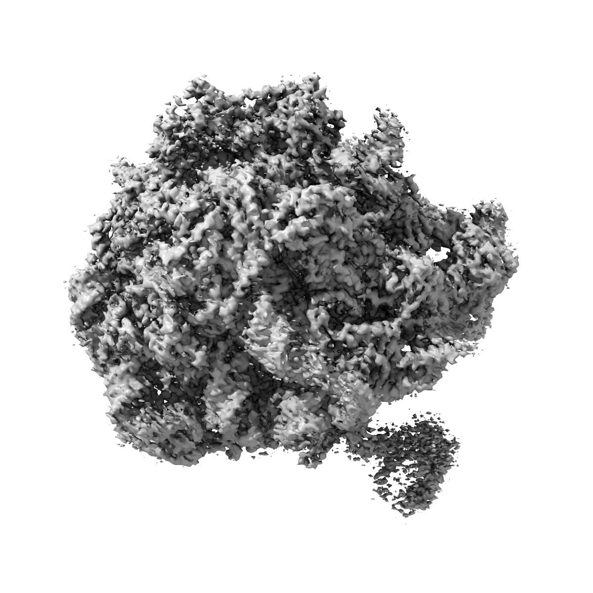
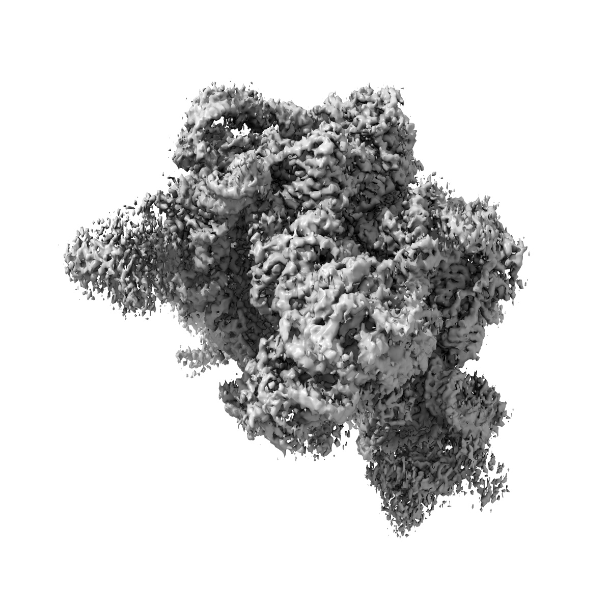
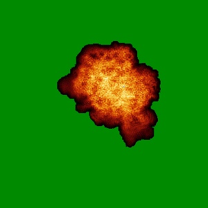
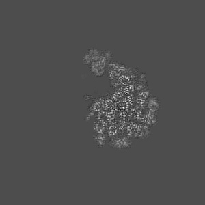
Cryo-EM reconstruction of the large subunit of the chloroplast ribosome
EMD-3531
Single-particle3.2 Å
 Deposition: 10/12/2016
Map released: 11/01/2017
Last modified: 15/05/2024
Concentration: 0.12
mg/mL
Buffer
pH: 7.6
Buffer components [6]:
Buffer components [6]:
| Name | Formula | Concentration | ChEBI |
|---|---|---|---|
| Tris hydrochloride | Tris-HCl | 25.0 mM | |
| Potassium chloride | KCl | 25.0 mM | |
| Magnesium acetate | Mg(CH3COO)2 | 25.0 mM | |
| Dithiothreitol | DTT | 2.0 mM | |
| Spermine | - | 0.05 mM | - |
| Spermidine | - | 2.0 mM | - |
Grid
Model:
Quantifoil R2/2
Material: COPPER
Material: COPPER
Pretreatment
Support Film [1]
| Material | Topology | Thickness |
|---|---|---|
| CARBON | CONTINUOUS | - |
Vitrification
Cryogen name: ETHANE-PROPANE
Chamber humidity: 100%
Chamber temperature: 278 K
Instrument: FEI VITROBOT MARK I
Chamber humidity: 100%
Chamber temperature: 278 K
Instrument: FEI VITROBOT MARK I
Microscope: FEI TITAN KRIOS
Illumination mode: FLOOD BEAM
Imaging mode: BRIGHT FIELD
Electron source: FIELD EMISSION GUN
Acceleration voltage: 300 kV
Nominal CS: 2.7 mm
Nominal defocus: 0.8 µm - 3.5 µm
Nominal magnification: 59000.0
Specimen holder model: FEI TITAN KRIOS AUTOGRID HOLDER
Illumination mode: FLOOD BEAM
Imaging mode: BRIGHT FIELD
Electron source: FIELD EMISSION GUN
Acceleration voltage: 300 kV
Nominal CS: 2.7 mm
Nominal defocus: 0.8 µm - 3.5 µm
Nominal magnification: 59000.0
Specimen holder model: FEI TITAN KRIOS AUTOGRID HOLDER
Image Recording
[1]
Detector model:
FEI FALCON II (4k x 4k)
Detector mode: INTEGRATING
Frames per image: 2-8
Number of real images: 2796
Average electron dose per image: 20.0 e/Å2
Detector mode: INTEGRATING
Frames per image: 2-8
Number of real images: 2796
Average electron dose per image: 20.0 e/Å2
Final
reconstruction
Resolution: 3.2
Å
(
BY AUTHOR)
Resolution method: FSC 0.143 CUT-OFF
Number of images used: 154332
Resolution method: FSC 0.143 CUT-OFF
Number of images used: 154332
⌯ Applied Symmetry
Point group:
C1
Software
[1]
| Name | Version | Details |
|---|---|---|
| RELION | 1.4 | - |
⦨ Initial angle
assignment
⦩ Final angle assignment
Particle selection
[1]
| Selected | Ref. model | Method | Software | Details |
|---|---|---|---|---|
| 326094 | - | - | - | - |
Final 3D classification
Software
[1]
| Name | Version | Details |
|---|---|---|
| RELION | 1.4 | - |
Format: CCP4
Data type: IMAGE STORED AS FLOATING POINT NUMBER (4 BYTES)
Annotation details: Cryo-EM reconstruction of the 50S large subunit of the chloroplast ribosome
Data type: IMAGE STORED AS FLOATING POINT NUMBER (4 BYTES)
Annotation details: Cryo-EM reconstruction of the 50S large subunit of the chloroplast ribosome
⬡ Geometry
| X | Y | Z | |
|---|---|---|---|
| Dimensions | 320 | 320 | 320 |
| Origin | 0 | 0 | 0 |
| Spacing | 320 | 320 | 320 |
| Voxel size | 1.39 Å | 1.39 Å | 1.39 Å |
Contour list
| Primary | Level | Source |
|---|---|---|
| True | 0.08 | AUTHOR |