EMD-25533

Single-particle
4.8 Å
EMD-25533 Deposition: 25/11/2021
Map released: 13/07/2022
Last modified: 28/02/2024
Overview 3D View Sample Experiment Validation Additional data Links

EMD-25533

Archive Files (Depositor)

Primary 3D volume (map.gz) Half-map 1 Half-map 2 (map.gz) Experimental metadata (xml) Experimental metadata (cif.gz)

Validation (wwPDB)

Map-only validation report (pdf.gz) Map-7sym summary report (pdf.gz) Map-7sym FULL report (pdf.gz)

EMDB Annotations

EMICSS entry mapping (xml)

EMDB Files

VA raw map (map) VA resolution mask (mrc)

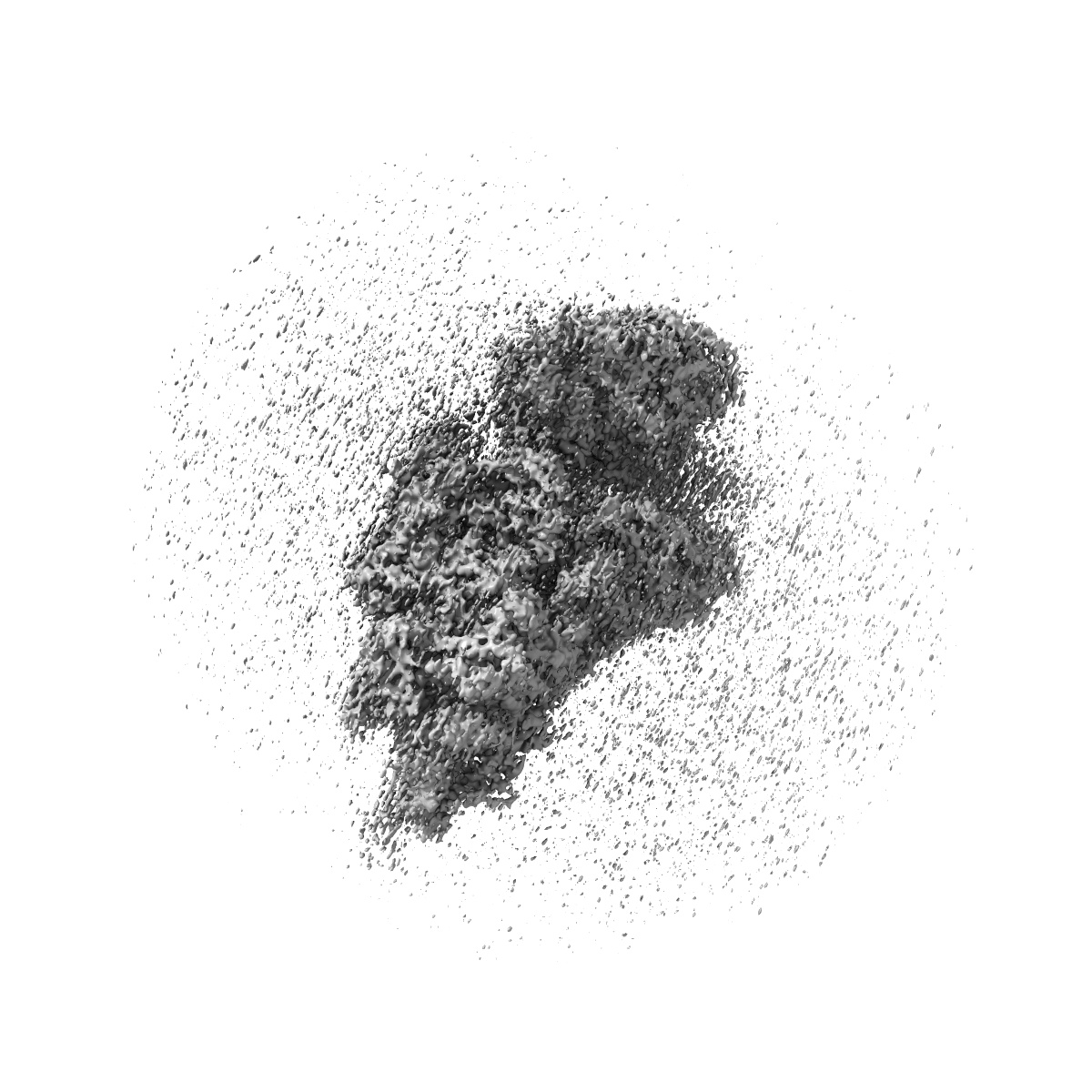
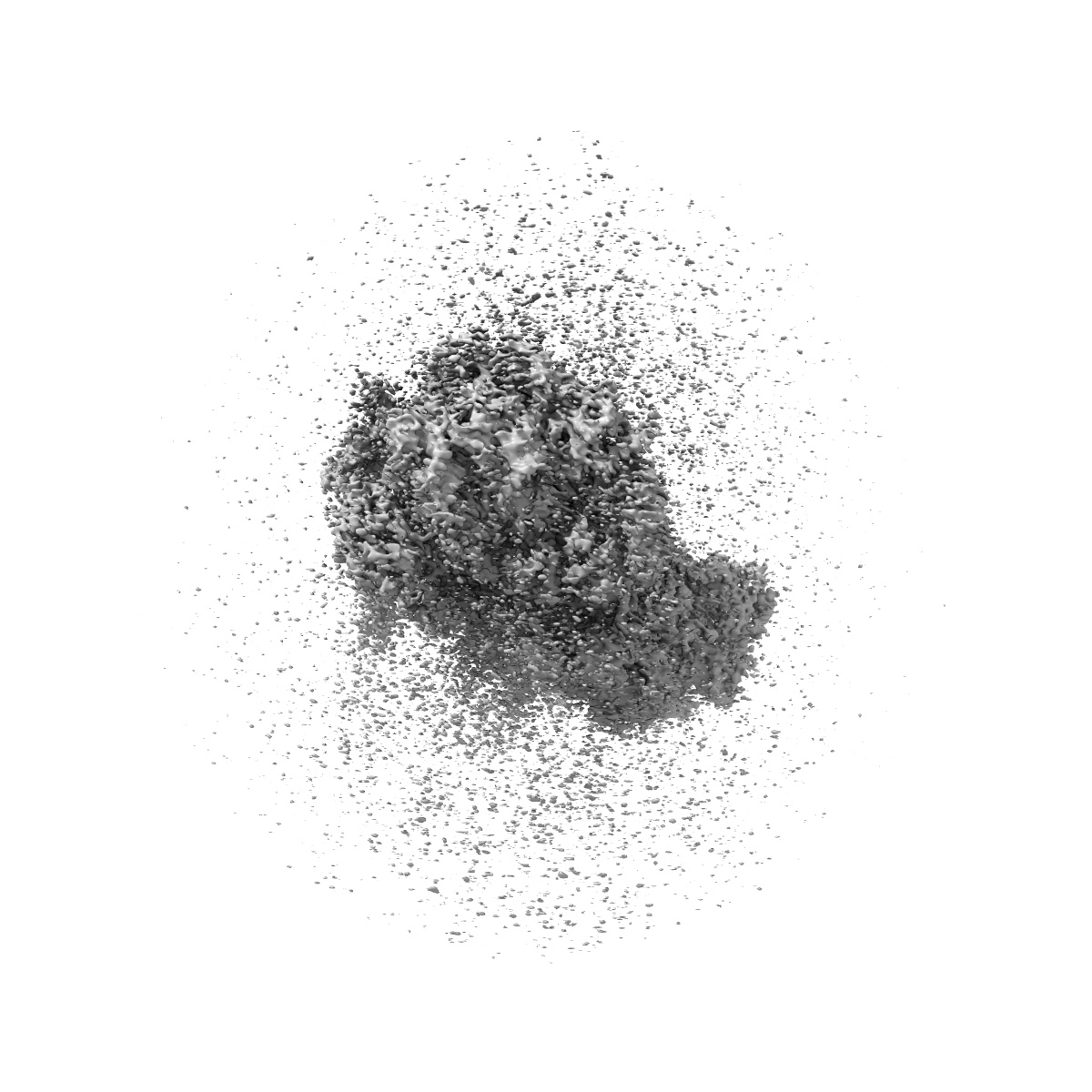
Structure of the HCV IRES bound to the 40S ribosomal subunit, head opening. Structure 7(delta dII)

EMD-25533

Single-particle
4.8 Å
EMD-25533 Deposition: 25/11/2021
Map released: 13/07/2022
Last modified: 28/02/2024
Overview 3D View Sample Experiment Validation Additional data Links
Sample Organism: Oryctolagus cuniculus, Hepatitis C virus (isolate 1)
Sample: 40S ribosomal small subunit with HCV IRES
Fitted models: 7sym

Deposition Authors: Brown ZP , Abaeva IS , De S , Hellen CUT , Pestova TV , Frank J

Comprehensive structural overview of the HCV IRES-mediated translation initiation pathway

Brown ZP , Abaeva IS , De S , Hellen CUT , Pestova TV , Frank J
To Be Published