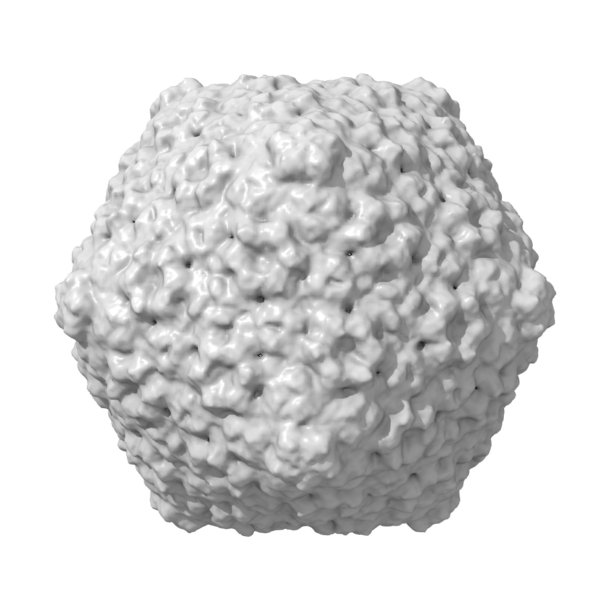
EMD-2133

Single-particle
15.0 Å
EMD-2133 Deposition: 11/06/2012
Map released: 21/11/2012
Last modified: 09/01/2013
Overview 3D View Sample Experiment Validation Additional data Links

EMD-2133

Archive Files (Depositor)

Primary 3D volume (map.gz) Half-map 1 Half-map 2 (map.gz) Experimental metadata (xml) Experimental metadata (cif.gz)

Validation (wwPDB)

Map-only validation report (pdf.gz)

EMDB Annotations

EMICSS entry mapping (xml)

The Structure of Lactococcal Phage TP901-1 by electron microscopy: the capsid

EMD-2133

Single-particle
15.0 Å
EMD-2133 Deposition: 11/06/2012
Map released: 21/11/2012
Last modified: 09/01/2013
Overview 3D View Sample Experiment Validation Additional data Links
Sample Organism: Lactococcus phage TP901-1
Sample: Icosahedral capsid of the lactococcal phage TP901-1

Deposition Authors: Bebeacua C, Lai L, Skovgaard Vegge C, Brondsted L , van Heel M, Veesler D, Cambillau C
Visualizing a complete Siphoviridae member by single-particle electron microscopy: the structure of lactococcal phage TP901-1.
Bebeacua C, Lai L, Skovgaard Vegge C, Brondsted L , van Heel M, Veesler D, Cambillau C
(2013) J.Virol. , 87 , 1061 - 1068