EMD-20762
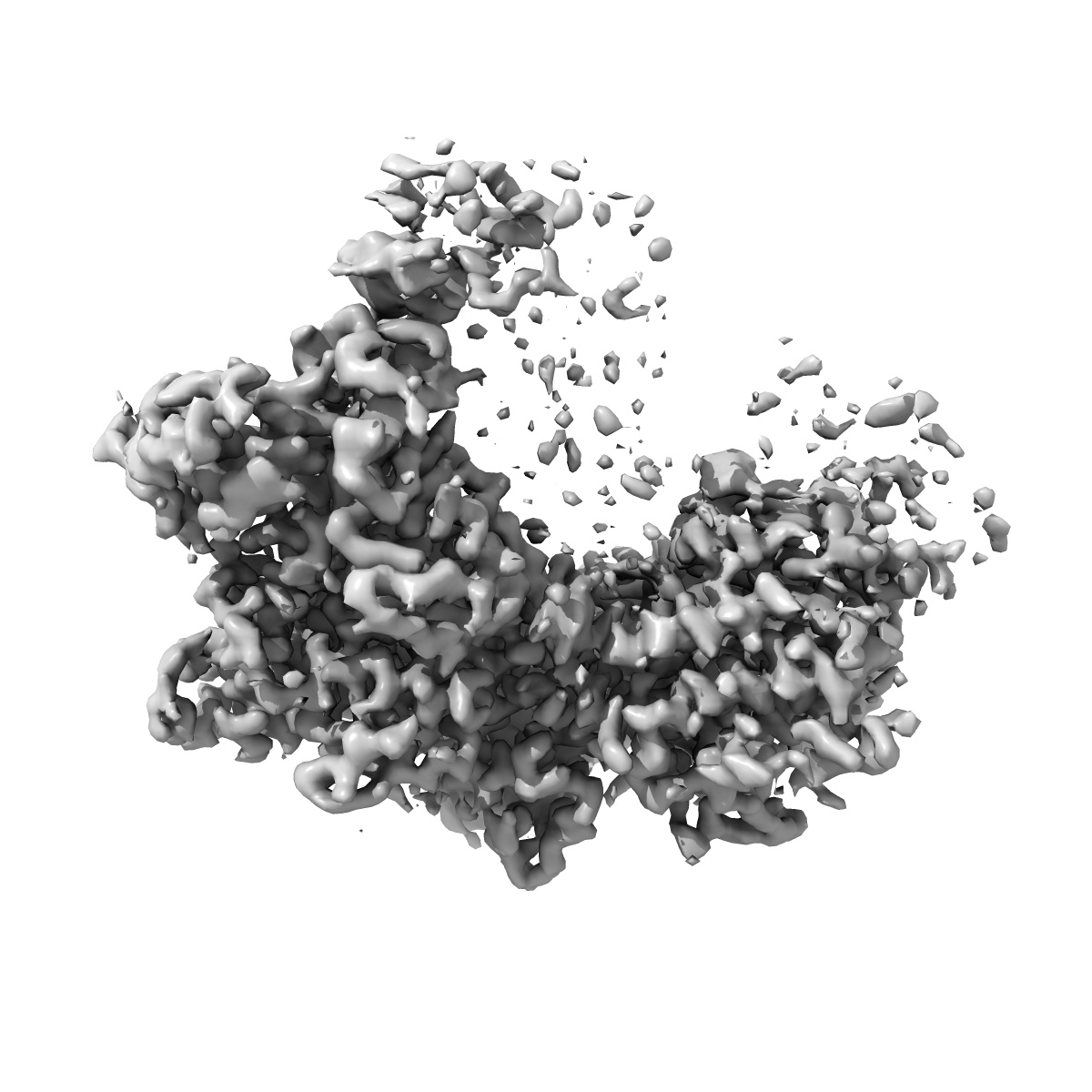
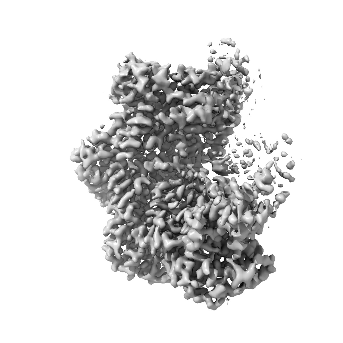
Katanin hexamer in the ring conformation in complex with substrate
EMD-20762
Single-particle3.6 Å
 Deposition: 26/09/2019
Map released: 09/10/2019
Last modified: 20/03/2024
Concentration: 1
mg/mL
Buffer
pH: 7.5
Buffer components [5]:
Buffer components [5]:
| Name | Formula | Concentration | ChEBI |
|---|---|---|---|
| HEPES | C8H18N2O4S | 20.0 mM | |
| Potassium chloride | KCl | 300.0 mM | |
| Magnesium dichloride | MgCl2 | 10.0 mM | |
| TCEP | C9H15O6P | 2.0 mM | |
| ATP | C10H16N5O13P3 | 1.0 mM |
Grid
Vitrification
Cryogen name: ETHANE
Chamber humidity: 90%
Chamber temperature: 279.15 K
Instrument: LEICA EM GP
Details: Load 5 ul sample, wait 10 sec, blot 4.5 sec.
Chamber humidity: 90%
Chamber temperature: 279.15 K
Instrument: LEICA EM GP
Details: Load 5 ul sample, wait 10 sec, blot 4.5 sec.
Microscope: FEI TITAN KRIOS
Illumination mode: FLOOD BEAM
Imaging mode: BRIGHT FIELD
Electron source: FIELD EMISSION GUN
Acceleration voltage: 300 kV
C2 aperture diameter: 70.0 µm
Nominal CS: 2.7 mm
Nominal defocus: 1.0 µm - 2.8000000000000003 µm
Calibrated defocus: 1.0 µm - 2.8000000000000003 µm
Nominal magnification: 130000.0
Specimen holder model: FEI TITAN KRIOS AUTOGRID HOLDER
Cooling holder cryogen: NITROGEN
Alignment procedure: COMA FREE
Temperature
Minimum: 93.0
K
Maximum: 103.0 K
Maximum: 103.0 K
Specialist optics
Energy filter
Image Recording
[1]
Detector model:
GATAN K2 SUMMIT (4k x 4k)
Detector mode: SUPER-RESOLUTION
Dimensions: 7420 pixel x 7676 pixel
Frames per image: 1-50
Number of grids: 4
Number of real images: 5911
Average exposure time: 10.0 s
Average electron dose per image: 73.0 e/Å2
Detector mode: SUPER-RESOLUTION
Dimensions: 7420 pixel x 7676 pixel
Frames per image: 1-50
Number of grids: 4
Number of real images: 5911
Average exposure time: 10.0 s
Average electron dose per image: 73.0 e/Å2
Details: Frames were aligned, dose weighted and summed using MotionCor2
Final
reconstruction
Resolution: 3.6
Å
(
BY AUTHOR)
Resolution method: FSC 0.143 CUT-OFF
Number of classed used: 1
Number of images used: 108700
Algorithm: FOURIER SPACE
Details:
Resolution method: FSC 0.143 CUT-OFF
Number of classed used: 1
Number of images used: 108700
Algorithm: FOURIER SPACE
Details:
⌯ Applied Symmetry
Point group:
C1
Software
[1]
| Name | Version | Details |
|---|---|---|
| RELION | 3.0 | - |
⦨ Initial angle
assignment
⦩ Final angle assignment
Particle selection
[1]
| Selected | Ref. model | Method | Software | Details |
|---|---|---|---|---|
| 1876223 | - | - | - | Particles were automatically picked |
Final 3D classification
Number of classes:
10
Avg. number of members per classes: 122136.0
Avg. number of members per classes: 122136.0
Software
[1]
| Name | Version | Details |
|---|---|---|
| RELION | 3.0 | - |
Format: CCP4
Data type: IMAGE STORED AS FLOATING POINT NUMBER (4 BYTES)
Annotation details: Katanin hexamer in the ring conformation in complex with substrate
Data type: IMAGE STORED AS FLOATING POINT NUMBER (4 BYTES)
Annotation details: Katanin hexamer in the ring conformation in complex with substrate
⬡ Geometry
| X | Y | Z | |
|---|---|---|---|
| Dimensions | 224 | 224 | 224 |
| Origin | 0 | 0 | 0 |
| Spacing | 224 | 224 | 224 |
| Voxel size | 1.08 Å | 1.08 Å | 1.08 Å |
Contour list
| Primary | Level | Source |
|---|---|---|
| True | 5.74 | AUTHOR |