represent EMBL in Europe.
Tissue biology and disease modelling
Structural biology
Main laboratory
EMBL-EBI: European Bioinformatics Institute
Epigenetics and neurobiology
Examples: 1001, Apoferritin, Tomography, Rossmann MG, 5A1A
advanced search
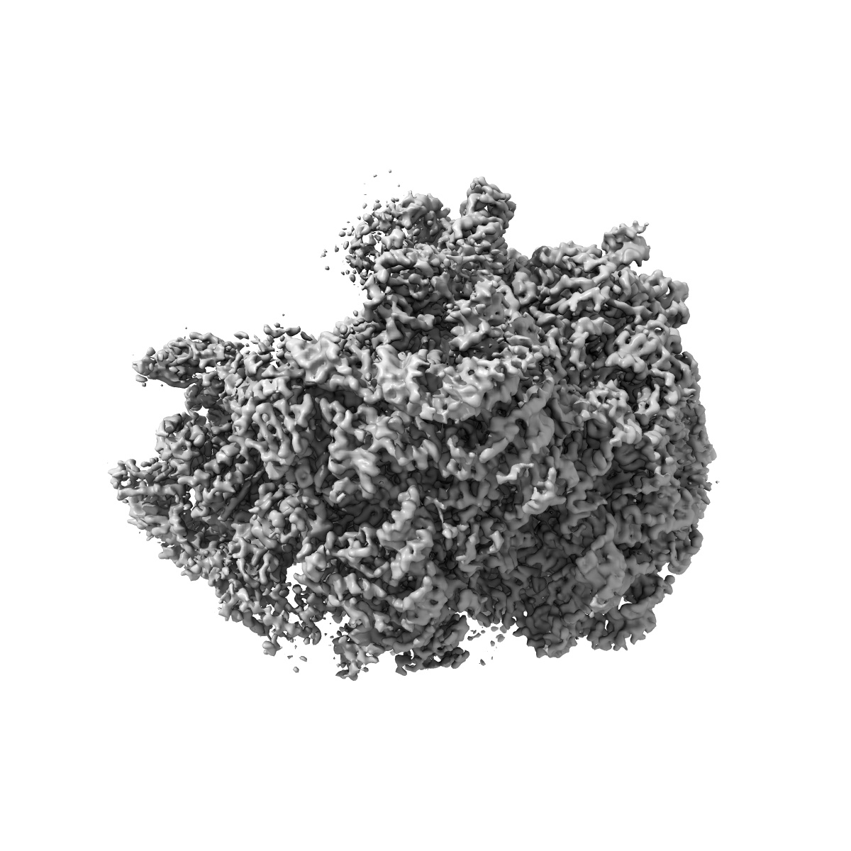
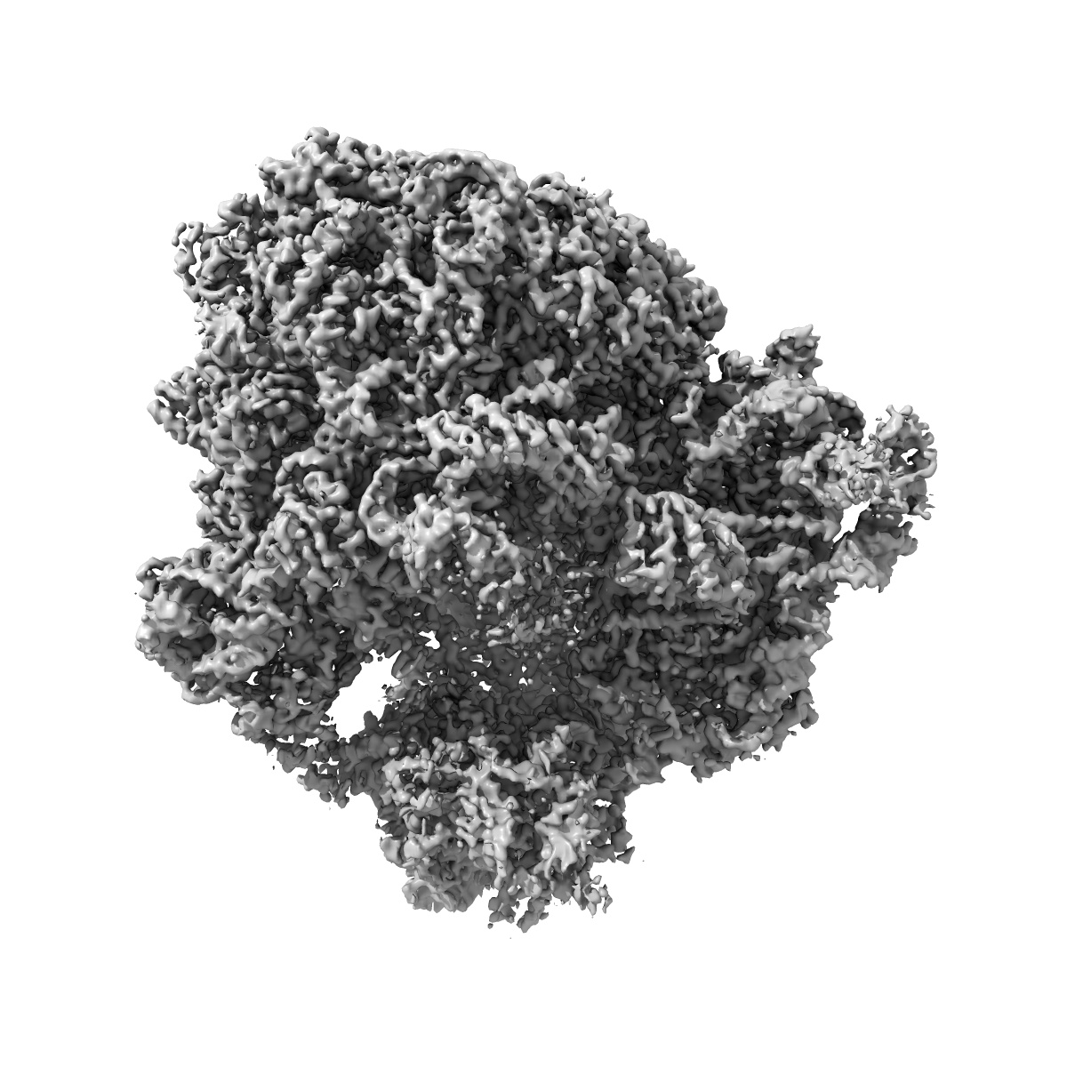
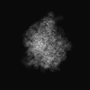
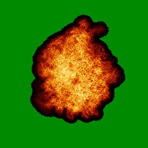
Structures of ribosome bound initiation factor 2 reveal the mechanism of subunit association
Searching in EMDBHelp
Share EMDB
 Deposition: 17/12/2015