EMD-45563

Helical reconstruction
2.8 Å
EMD-45563 Deposition: 27/06/2024
Map released: 12/02/2025
Last modified: 12/03/2025
Overview 3D View Sample Experiment Validation Additional data Links

EMD-45563

Archive Files (Depositor)

Primary 3D volume (map.gz) Half-map 1 Half-map 2 (map.gz) Experimental metadata (xml) Experimental metadata (cif.gz)

Validation (wwPDB)

Map-only validation report (pdf.gz) Map-9cfu summary report (pdf.gz) Map-9cfu FULL report (pdf.gz)

EMDB Annotations

EMICSS entry mapping (xml)

EMDB Files

VA raw map (map) VA resolution mask (mrc)

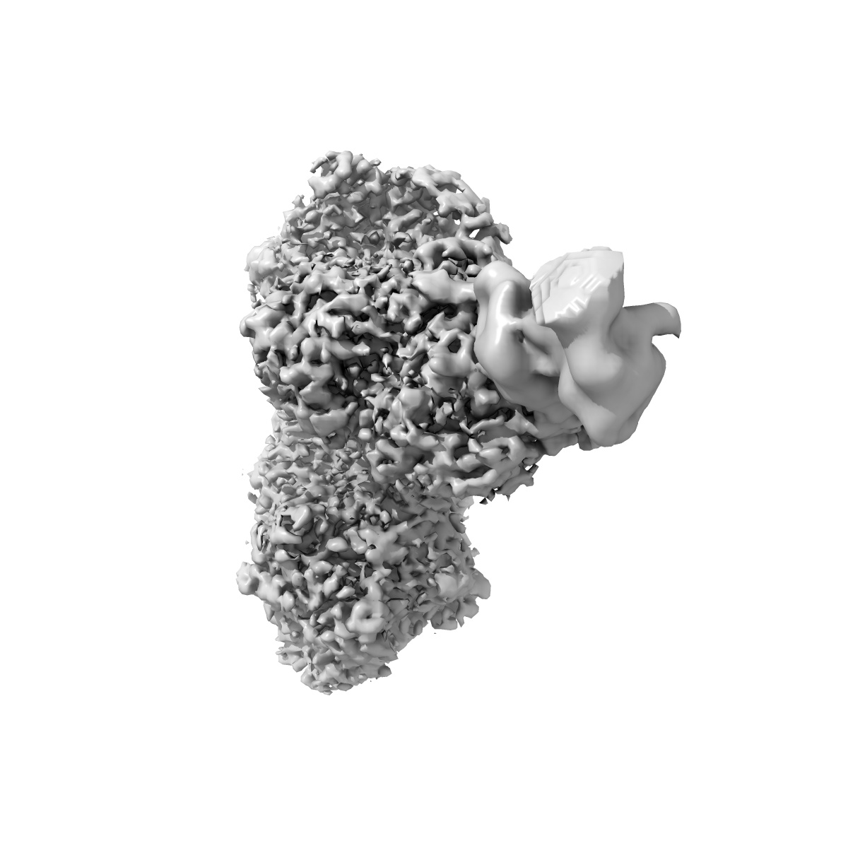
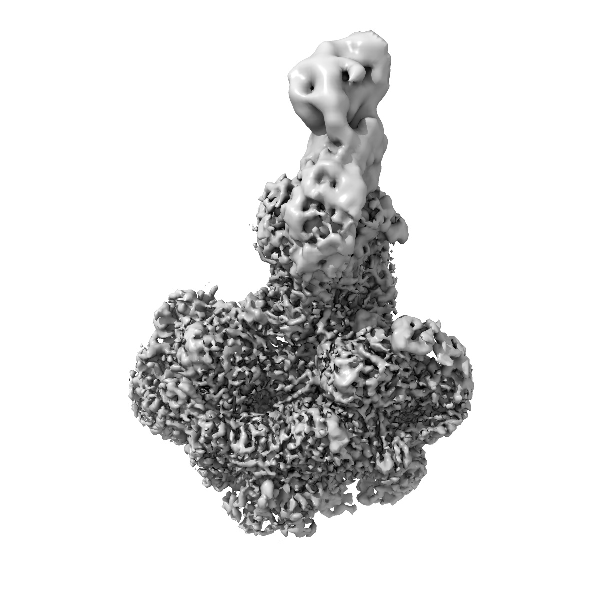
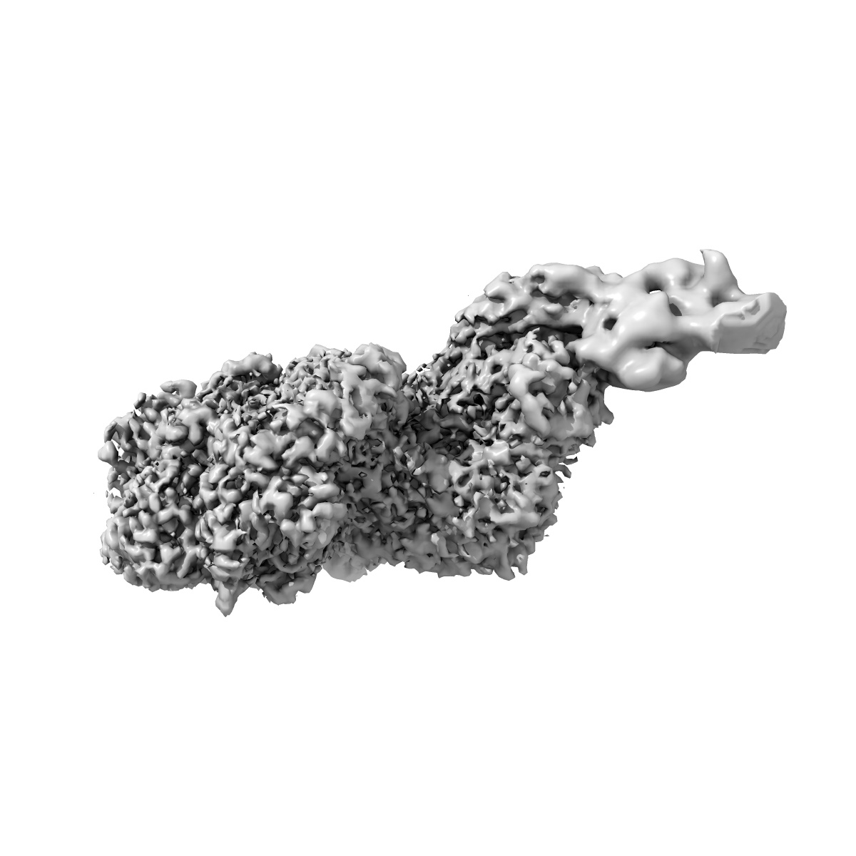
Cryo-EM structure of myosin-1c bound to F-actin in the ADP-A state

EMD-45563

Helical reconstruction
2.8 Å
EMD-45563 Deposition: 27/06/2024
Map released: 12/02/2025
Last modified: 12/03/2025
Overview 3D View Sample Experiment Validation Additional data Links
Sample Organism: Mus musculus, Rattus norvegicus, Oryctolagus cuniculus
Sample: Complex of myosin-1c with F-actin
Fitted models: 9cfu

Deposition Authors: Chavali SS, Sindelar CV , Ostap ME
High-resolution structures of Myosin-IC reveal a unique actin-binding orientation, ADP release pathway, and power stroke trajectory.
Chavali SS, Carman PJ, Shuman H , Ostap EM , Sindelar CV
(2025) PNAS , 122 , e2415457122 - e2415457122
PUBMED: 40014570
DOI: doi:10.1073/pnas.2415457122
ISSN: 1091-6490
ASTM: PNASA6