EMD-44135

Single-particle
3.41 Å
EMD-44135 Deposition: 19/03/2024
Map released: 29/05/2024
Last modified: 21/08/2024
Overview 3D View Sample Experiment Validation Additional data Links

EMD-44135

Archive Files (Depositor)

Primary 3D volume (map.gz) Half-map 1 Half-map 2 (map.gz) Experimental metadata (xml) Experimental metadata (cif.gz)

Validation (wwPDB)

Map-only validation report (pdf.gz) Map-9b3d summary report (pdf.gz) Map-9b3d FULL report (pdf.gz)

EMDB Annotations

EMICSS entry mapping (xml)

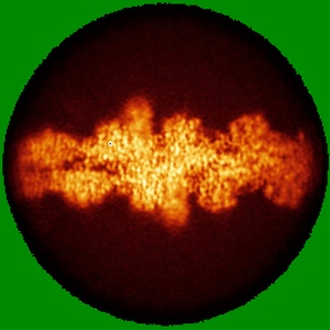
Dia1 in the Middle of F-actin

EMD-44135

Single-particle
3.41 Å
EMD-44135 Deposition: 19/03/2024
Map released: 29/05/2024
Last modified: 21/08/2024
Overview 3D View Sample Experiment Validation Additional data Links
Sample Organism: Oryctolagus cuniculus, Mus musculus
Sample: Structure of mDia1 dimer bound at the barbed end of actin filaments.
Fitted models: 9b3d

Deposition Authors: Palmer NJ , Barrie KR , Dominguez R
Mechanisms of actin filament severing and elongation by formins.
Palmer NJ , Barrie KR , Dominguez R
(2024) Nature , 632 , 437 - 442
PUBMED: 38843827
DOI: doi:10.1038/s41586-024-07637-0
ISSN: 1476-4687
ASTM: NATUAS