EMD-25468

Electron Crystallography
EMD-25468 Deposition: 19/11/2021
Map released: 07/09/2022
Last modified: 23/10/2024
Overview 3D View Sample Experiment Validation Additional data Links

EMD-25468

Archive Files (Depositor)

Primary 3D volume (map.gz) Half-map 1 Half-map 2 (map.gz) Experimental metadata (xml) Experimental metadata (cif.gz)

Validation (wwPDB)

Map-only validation report (pdf.gz) Map-7swa summary report (pdf.gz) Map-7swa FULL report (pdf.gz)

EMDB Annotations

EMICSS entry mapping (xml)

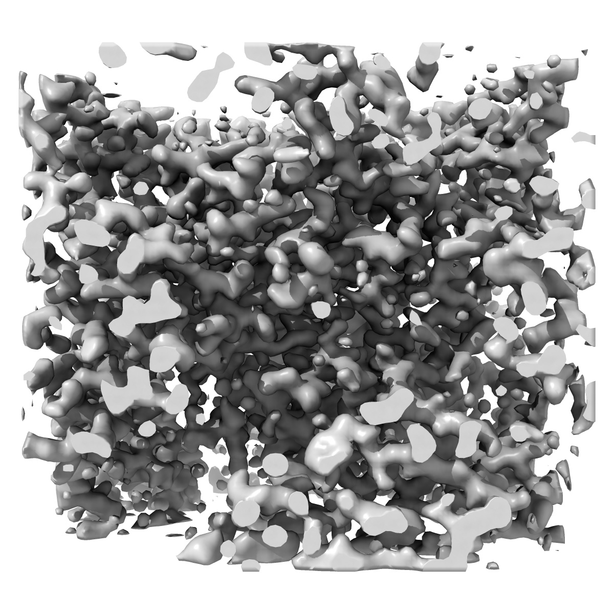
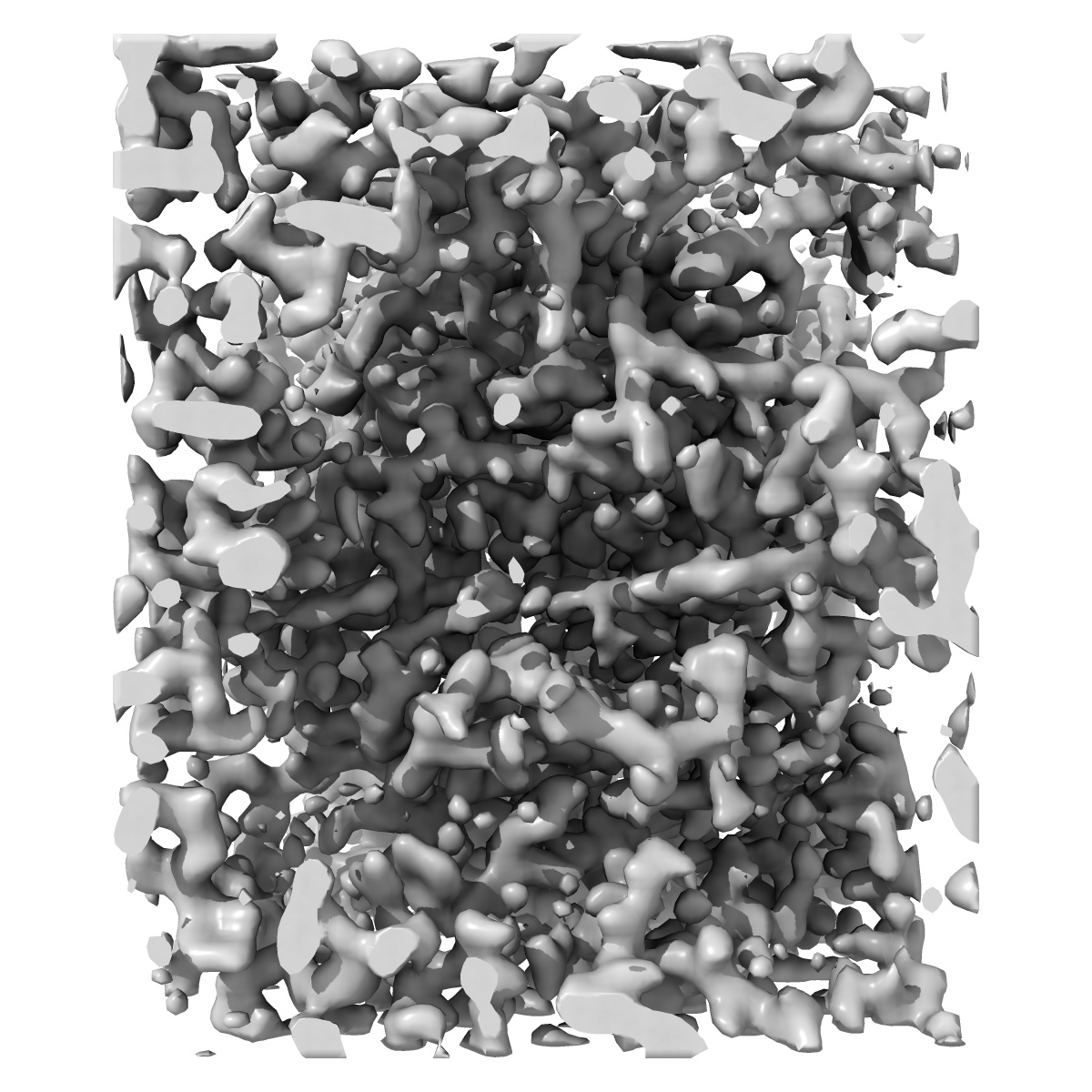
MicroED structure of proteinase K from a 320 nm thick lamella measured at 300 kV

EMD-25468

Electron Crystallography
EMD-25468 Deposition: 19/11/2021
Map released: 07/09/2022
Last modified: 23/10/2024
Overview 3D View Sample Experiment Validation Additional data Links
Sample Organism: Parengyodontium album
Sample: Proteinase K
Fitted models: 7swa

Deposition Authors: Martynowycz MW , Clabbers MTB
Benchmarking the ideal sample thickness in cryo-EM.
Martynowycz MW , Clabbers MTB , Unge J , Hattne J , Gonen T
(2021) PNAS , 118
PUBMED: 34873060
DOI: doi:10.1073/pnas.2108884118
ISSN: 1091-6490
ASTM: PNASA6