EMD-14355

Helical reconstruction
3.7 Å
EMD-14355 Deposition: 15/02/2022
Map released: 10/08/2022
Last modified: 09/07/2025
Overview 3D View Sample Experiment Validation Additional data Links

EMD-14355

Archive Files (Depositor)

Primary 3D volume (map.gz) Half-map 1 Half-map 2 (map.gz) Experimental metadata (xml) Experimental metadata (cif.gz)

Validation (wwPDB)

Map-only validation report (pdf.gz) Map-7yx5 summary report (pdf.gz) Map-7yx5 FULL report (pdf.gz)

EMDB Annotations

EMICSS entry mapping (xml)

EMDB Files

VA raw map (map) VA resolution mask (mrc)

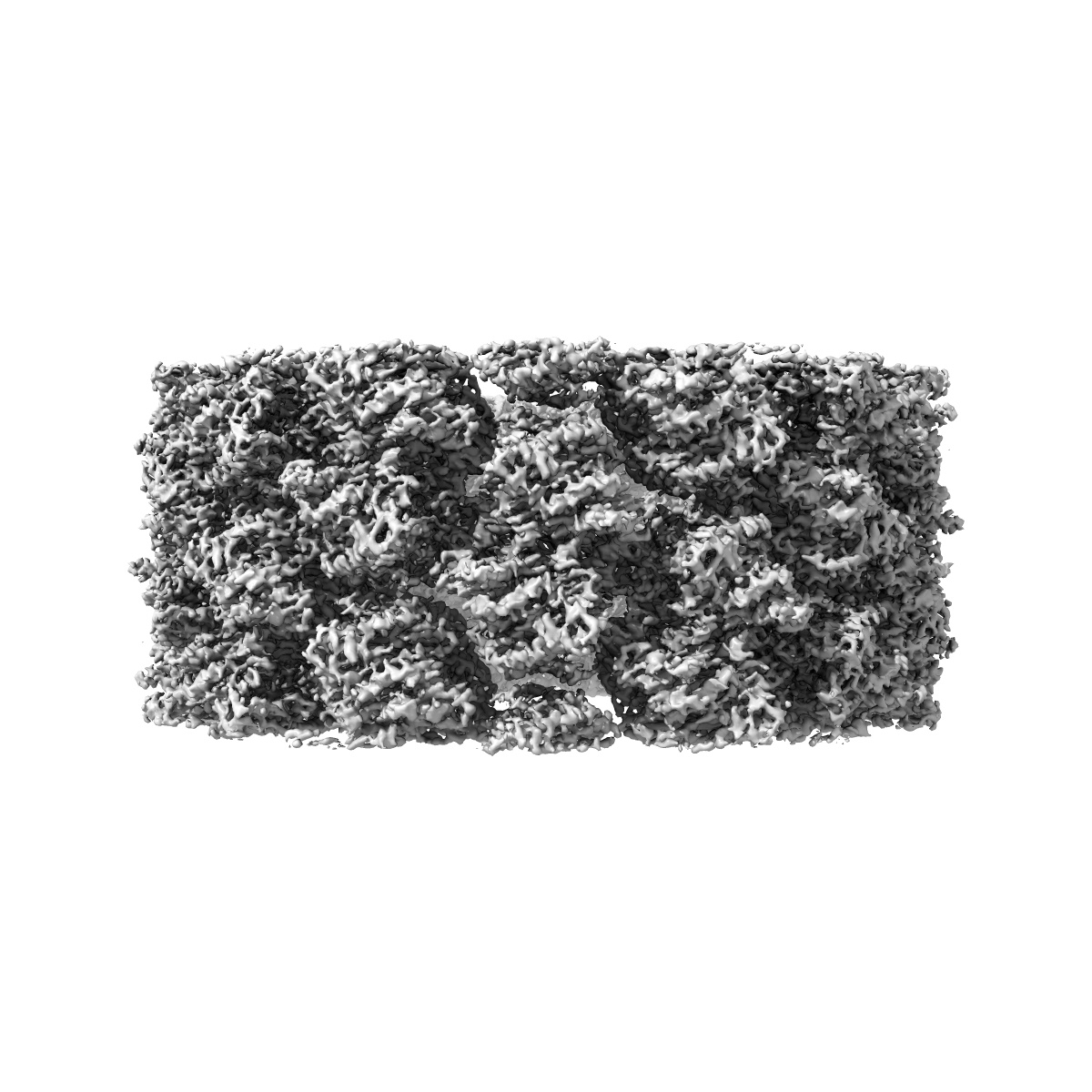
Structure of the Mimivirus genomic fibre in its relaxed 5-start helix form

EMD-14355

Helical reconstruction
3.7 Å
EMD-14355 Deposition: 15/02/2022
Map released: 10/08/2022
Last modified: 09/07/2025
Overview 3D View Sample Experiment Validation Additional data Links
Sample Organism: Acanthamoeba polyphaga mimivirus
Sample: Mimivirus genomic fibre in its relaxed 5-start helix form
Fitted models: 7yx5

Deposition Authors: Villalta A , Schmitt A
The giant mimivirus 1.2 Mb genome is elegantly organized into a 30 nm diameter helical protein shield.
PUBMED: 35900198
DOI: doi:10.7554/eLife.77607
ISSN: 2050-084X