









EMD-13641
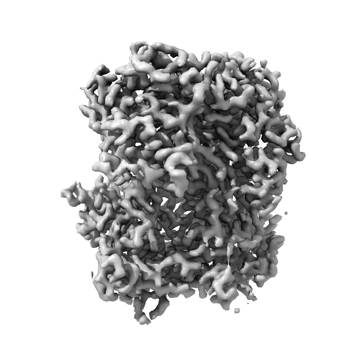
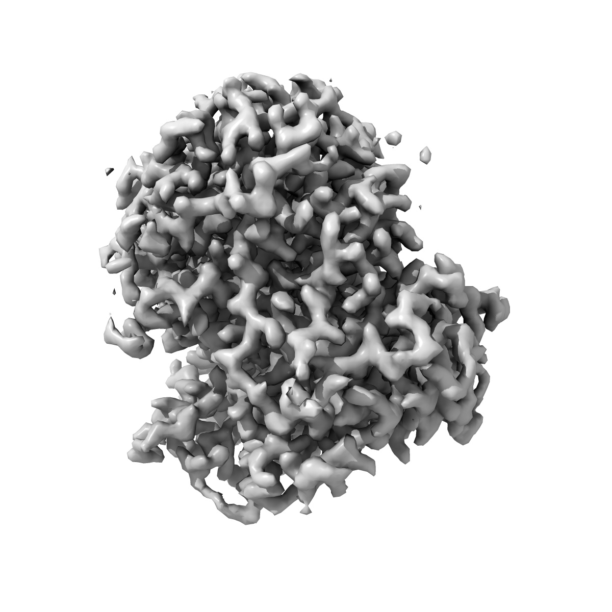
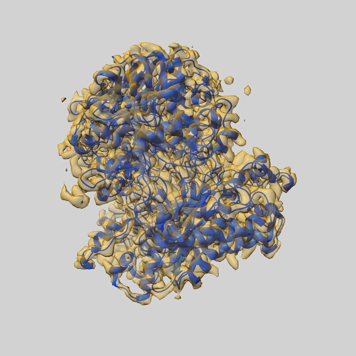
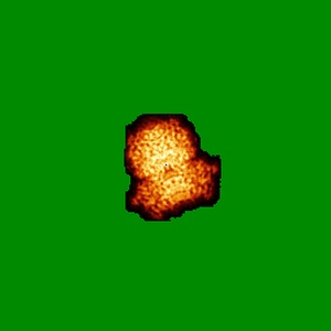
Structure of the Mimivirus genomic fibre asymmetric unit
EMD-13641
Single-particle3.3 Å
 Deposition: 27/09/2021
Map released: 10/08/2022
Last modified: 17/07/2024
Sample Organism:
Acanthamoeba polyphaga mimivirus
Sample: Structure of the Mimivirus genomic fibre asymmetric unit
Fitted models: 7ptv
Deposition Authors: Villalta A ,
Schmitt A ,
Estrozi LF ,
Quemin ERJ ,
Alempic JM,
Lartigue A,
Prazak V ,
Belmudes L,
Vasishtan D,
Colmant AMG ,
Honore FA ,
Coute Y ,
Grunewald K ,
Abergel C
,
Schmitt A ,
Estrozi LF ,
Quemin ERJ ,
Alempic JM,
Lartigue A,
Prazak V ,
Belmudes L,
Vasishtan D,
Colmant AMG ,
Honore FA ,
Coute Y ,
Grunewald K ,
Abergel C
Sample: Structure of the Mimivirus genomic fibre asymmetric unit
Fitted models: 7ptv
Deposition Authors: Villalta A
 ,
Schmitt A ,
Estrozi LF ,
Quemin ERJ ,
Alempic JM,
Lartigue A,
Prazak V ,
Belmudes L,
Vasishtan D,
Colmant AMG ,
Honore FA ,
Coute Y ,
Grunewald K ,
Abergel C
,
Schmitt A ,
Estrozi LF ,
Quemin ERJ ,
Alempic JM,
Lartigue A,
Prazak V ,
Belmudes L,
Vasishtan D,
Colmant AMG ,
Honore FA ,
Coute Y ,
Grunewald K ,
Abergel C
The giant mimivirus 1.2 Mb genome is elegantly organized into a 30 nm diameter helical protein shield.
Villalta A ,
Schmitt A ,
Estrozi LF ,
Quemin ERJ ,
Alempic JM,
Lartigue A,
Prazak V ,
Belmudes L,
Vasishtan D,
Colmant AMG ,
Honore FA ,
Coute Y ,
Grunewald K ,
Abergel C
(2022) eLife , 11
,
Schmitt A ,
Estrozi LF ,
Quemin ERJ ,
Alempic JM,
Lartigue A,
Prazak V ,
Belmudes L,
Vasishtan D,
Colmant AMG ,
Honore FA ,
Coute Y ,
Grunewald K ,
Abergel C
(2022) eLife , 11
Grant Support:
- European Research Council (ERC): 832601 (European Union)
- Agence Nationale de la Recherche (ANR): ANR-16-CE11-0033-01 (France)
- German Research Foundation (DFG): (INST 152/772-1|152/774-1|152/775-1|152/776-1 (Germany)
- Wellcome Trust: 107806/Z/15/Z (United Kingdom)
- German Federal Ministry for Education and Research: 05K18BHA (Germany)
- Alexander von Humboldt Foundation: FRA 1200789 HFST-P (Germany)
- Agence Nationale de la Recherche (ANR): ANR-10-INBS-04 (France)