EMD-10205

Single-particle
2.73 Å
EMD-10205 Deposition: 08/08/2019
Map released: 13/05/2020
Last modified: 22/05/2024
Overview 3D View Sample Experiment Validation Additional data Links

EMD-10205

Archive Files (Depositor)

Primary 3D volume (map.gz) Half-map 1 Half-map 2 (map.gz) Experimental metadata (xml) Experimental metadata (cif.gz)

Validation (wwPDB)

Map-only validation report (pdf.gz) Map-6sht summary report (pdf.gz) Map-6sht FULL report (pdf.gz)

EMDB Annotations

EMICSS entry mapping (xml)

EMDB Files

VA raw map (map) VA resolution mask (mrc)

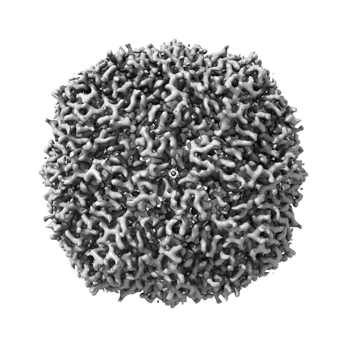
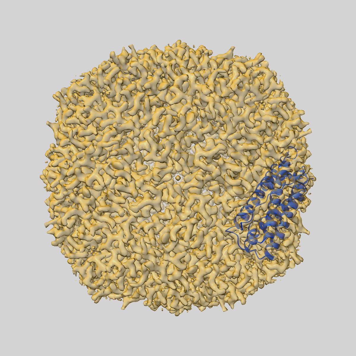
Molecular structure of mouse apoferritin resolved at 2.7 Angstroms with the Glacios cryo-microscope

EMD-10205

Single-particle
2.73 Å
EMD-10205 Deposition: 08/08/2019
Map released: 13/05/2020
Last modified: 22/05/2024
Overview 3D View Sample Experiment Validation Additional data Links
Sample Organism: Mus musculus
Sample: Mouse apoferritin
Fitted models: 6sht

Deposition Authors: Hamdi F , Tueting C
2.7 angstrom cryo-EM structure of vitrified M. musculus H-chain apoferritin from a compact 200 keV cryo-microscope.
Hamdi F , Tuting C , Semchonok DA , Visscher KM, Kyrilis FL , Meister A , Skalidis I , Schmidt L , Parthier C , Stubbs MT , Kastritis PL
(2020) PLoS One , 15 , e0232540 - e0232540
PUBMED: 32374767
DOI: doi:10.1371/journal.pone.0232540
ISSN: 1932-6203