EMD-0656

Single-particle
3.53 Å
EMD-0656 Deposition: 12/03/2019
Map released: 23/09/2020
Last modified: 20/03/2024
Overview 3D View Sample Experiment Validation Additional data Links

EMD-0656

Archive Files (Depositor)

Primary 3D volume (map.gz) Half-map 1 Half-map 2 (map.gz) Experimental metadata (xml) Experimental metadata (cif.gz)

Validation (wwPDB)

Map-only validation report (pdf.gz) Map-6o8w summary report (pdf.gz) Map-6o8w FULL report (pdf.gz)

EMDB Annotations

EMICSS entry mapping (xml)

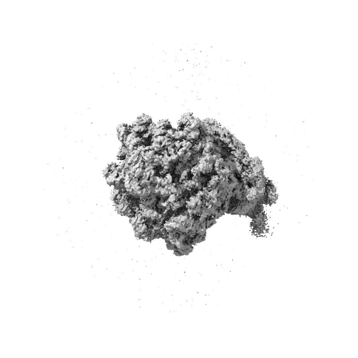
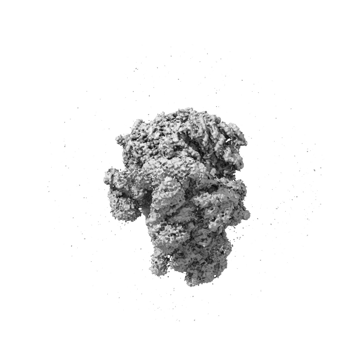
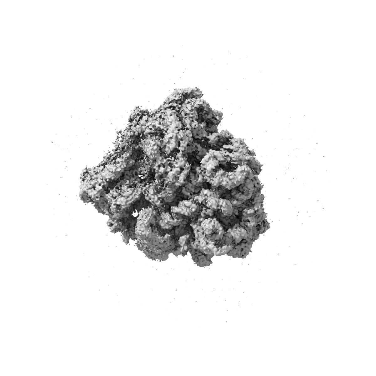
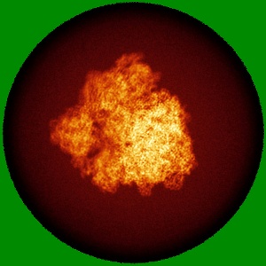
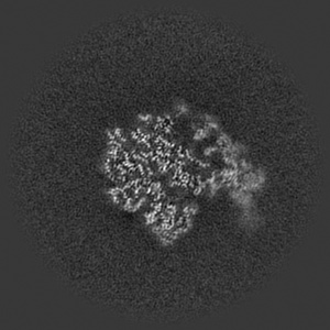
Cryo-EM image reconstruction of the 70S Ribosome Enterococcus faecalis Class01

EMD-0656

Single-particle
3.53 Å
EMD-0656 Deposition: 12/03/2019
Map released: 23/09/2020
Last modified: 20/03/2024
Overview 3D View Sample Experiment Validation Additional data Links
Sample Organism: Enterococcus faecalis
Sample: Enterococcus faecalis
Fitted models: 6o8w

Deposition Authors: Jogl G, Khayat R
Cryo-electron microscopy structure of the 70S ribosome from Enterococcus faecalis.
Murphy EL, Singh KV, Avila B , Kleffmann T, Gregory ST, Murray BE, Krause KL, Khayat R, Jogl G
(2020) Sci Rep , 10 , 16301 - 16301
PUBMED: 33004869
DOI: doi:10.1038/s41598-020-73199-6
ISSN: 2045-2322