EMD-0343

Electron Crystallography
2.75 Å
EMD-0343 Deposition: 20/11/2018
Map released: 06/02/2019
Last modified: 06/11/2024
Overview 3D View Sample Experiment Validation Additional data Links

EMD-0343

Archive Files (Depositor)

Primary 3D volume (map.gz) Half-map 1 Half-map 2 (map.gz) Experimental metadata (xml) Experimental metadata (cif.gz)

Validation (wwPDB)

Map-only validation report (pdf.gz) Map-6n4u summary report (pdf.gz) Map-6n4u FULL report (pdf.gz)

EMDB Annotations

EMICSS entry mapping (xml)

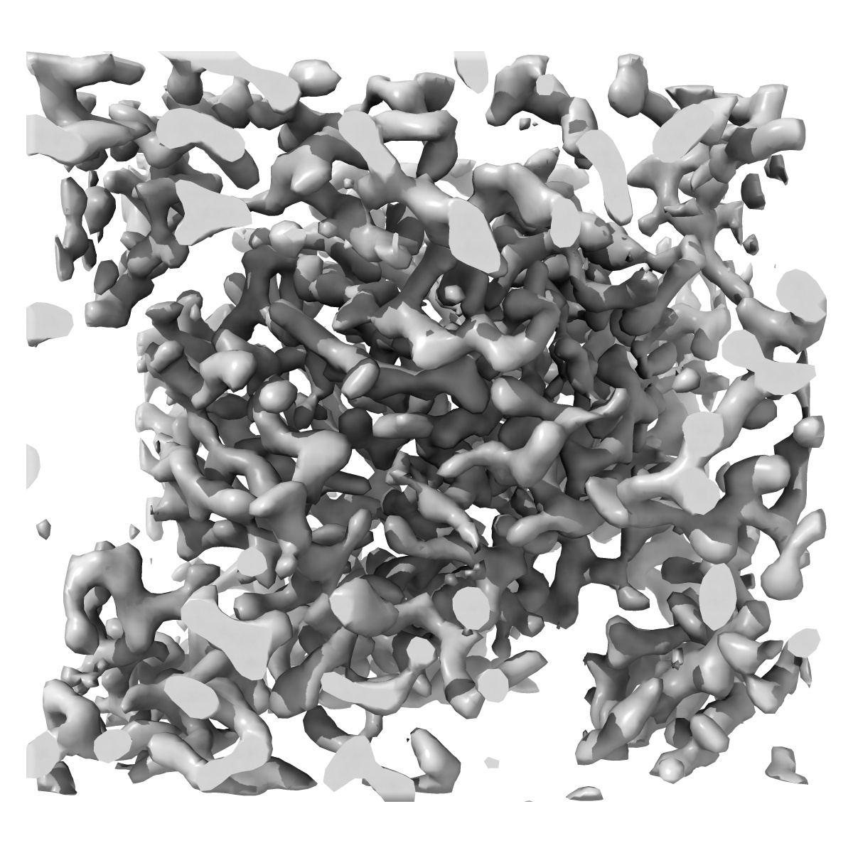
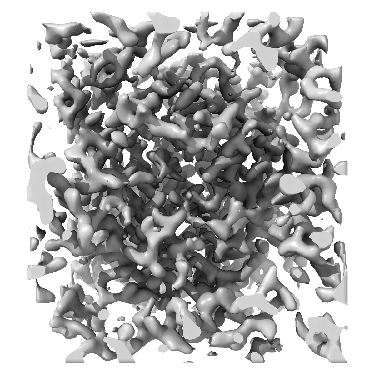
MicroED structure of Proteinase K at 2.75A resolution from a single milled crystal.

EMD-0343

Electron Crystallography
2.75 Å
EMD-0343 Deposition: 20/11/2018
Map released: 06/02/2019
Last modified: 06/11/2024
Overview 3D View Sample Experiment Validation Additional data Links
Sample Organism: Engyodontium album
Sample: Proteinase K
Fitted models: 6n4u

Deposition Authors: Martynowycz MW , Zhao W
Collection of Continuous Rotation MicroED Data from Ion Beam-Milled Crystals of Any Size.
Martynowycz MW , Zhao W, Hattne J , Jensen GJ, Gonen T
(2019) Structure , 27 , 545 - 548.e2
PUBMED: 30661853
DOI: doi:10.1016/j.str.2018.12.003
ISSN: 1878-4186
ASTM: STRUE6