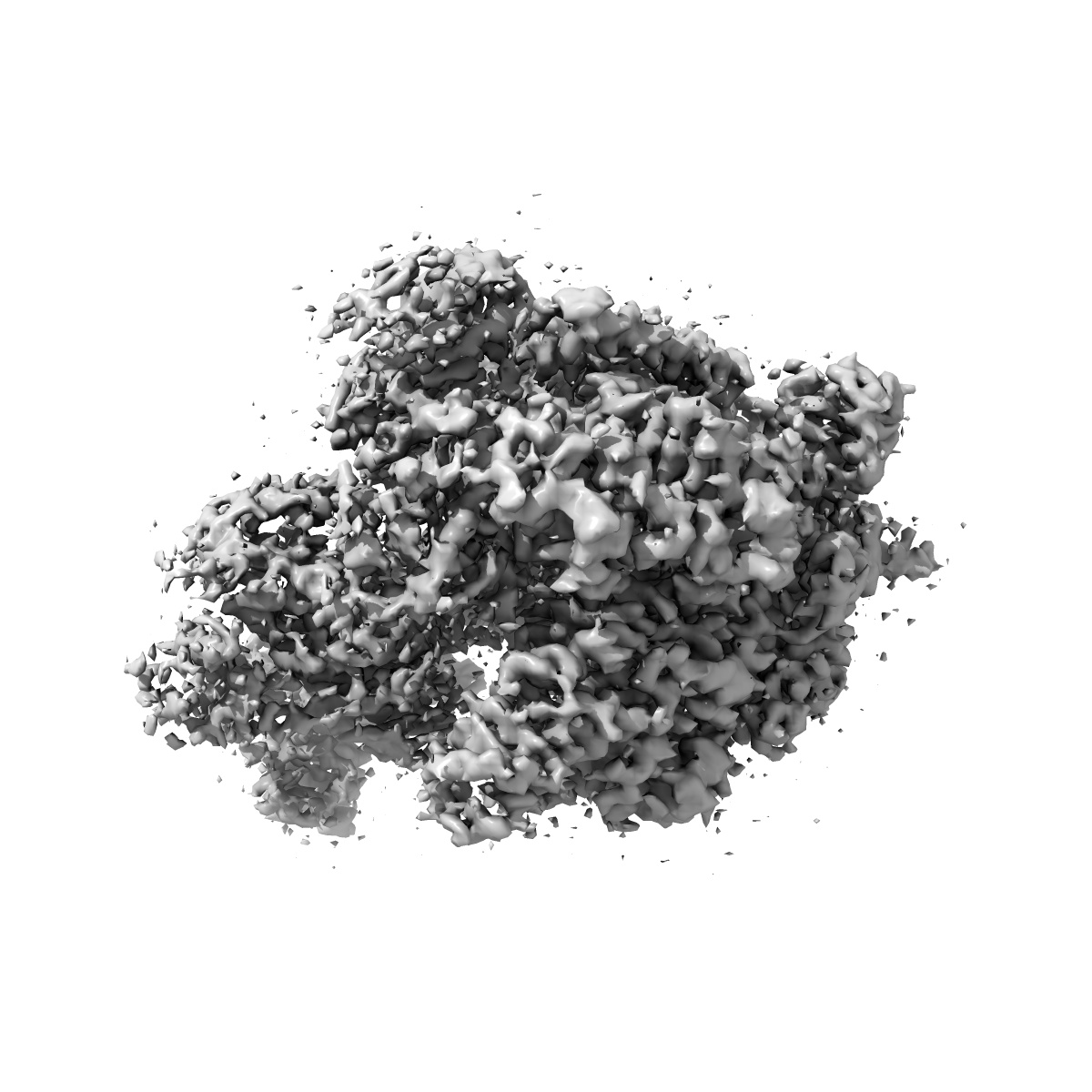
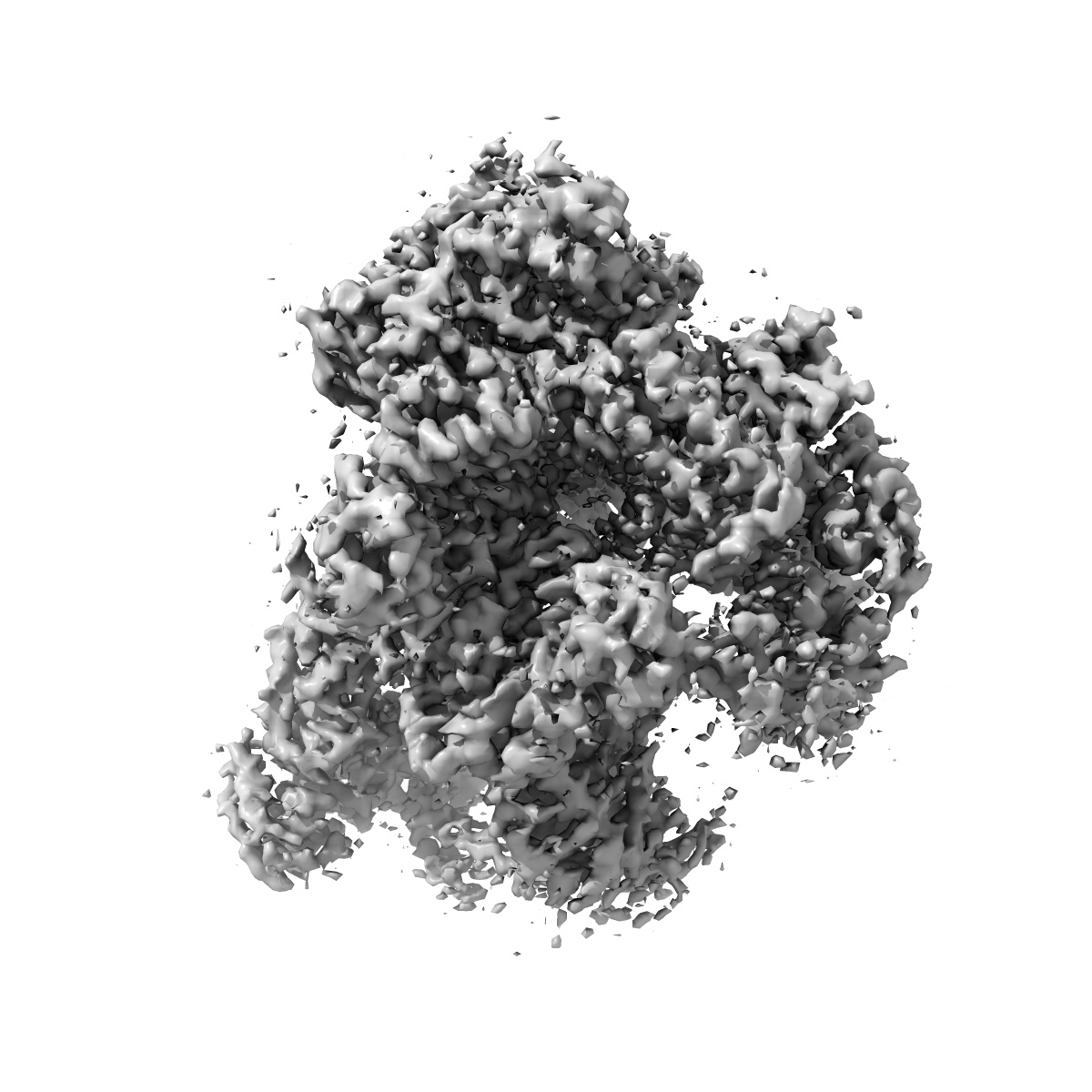
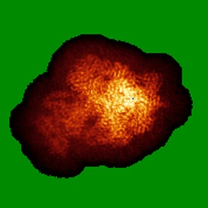
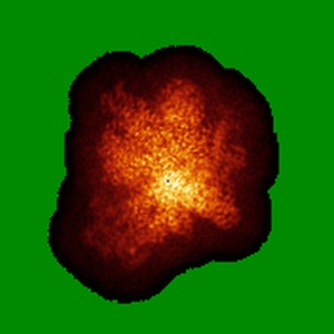
EMD-9961
Thermococcus kodakarensis RNA polymerase in complex with transcription factor E
EMD-9961
Single-particle4.0 Å
 Deposition: 03/07/2019
Map released: 01/07/2020
Last modified: 23/12/2020
Concentration: 0.3
mg/mL
Buffer
pH: 8.0
Grid
Vitrification
Cryogen name: ETHANE
Chamber humidity: 90%
Chamber temperature: 278 K
Instrument: FEI VITROBOT MARK IV
Chamber humidity: 90%
Chamber temperature: 278 K
Instrument: FEI VITROBOT MARK IV
Microscope: FEI TITAN KRIOS
Illumination mode: FLOOD BEAM
Imaging mode: BRIGHT FIELD
Electron source: FIELD EMISSION GUN
Acceleration voltage: 300 kV
C2 aperture diameter: 70.0 µm
Nominal CS: 0.002 mm
Nominal defocus: 1.5 µm - 3.5 µm
Nominal magnification: 47000.0
Calibrated magnification: 100000.0
Specimen holder model: FEI TITAN KRIOS AUTOGRID HOLDER
Cooling holder cryogen: NITROGEN
Alignment procedure: ZEMLIN TABLEAU
Specialist optics
SPH aberration_corrector:
Image Cs corrector
Image Recording
[1]
Detector model:
FEI FALCON II (4k x 4k)
Detector mode: INTEGRATING
Frames per image: 1-30
Number of real images: 2698
Average exposure time: 1.8 s
Average electron dose per image: 63.0 e/Å2
Detector mode: INTEGRATING
Frames per image: 1-30
Number of real images: 2698
Average exposure time: 1.8 s
Average electron dose per image: 63.0 e/Å2
Details: Movie frame data was subjected to motion correction to generate summed micrographs using MotionCor2.
Final
reconstruction
Resolution: 4.0
Å
(
BY AUTHOR)
Resolution method: FSC 0.143 CUT-OFF
Number of classed used: 1
Number of images used: 252508
Algorithm: BACK PROJECTION
Details:
Resolution method: FSC 0.143 CUT-OFF
Number of classed used: 1
Number of images used: 252508
Algorithm: BACK PROJECTION
Details:
⌯ Applied Symmetry
Point group:
C1
Software
[1]
| Name | Version | Details |
|---|---|---|
| RELION | 2.1 | - |
Startup model
[1]
⦨ Initial angle
assignment
⦩ Final angle assignment
Particle selection
[1]
| Selected | Ref. model | Method | Software | Details |
|---|---|---|---|---|
| 1226339 | - | - | - | Approximately 20,000 particles were selected manually. 2D class averages from the manual-picked particles were used as a template for automated particle picking in Relion 2.1. |
Final 3D classification
Number of classes:
8
Avg. number of members per classes: 51207.0
Avg. number of members per classes: 51207.0
Software
[1]
| Name | Version | Details |
|---|---|---|
| RELION | 2.1 | - |
CTF correction
Details: Defocus parameters were calculated from motion-corrected micrographs, using Gctf 1.06.
Using the defocus parameters, Cs (0.003, after Cs image corrector tuning) and amplitude contrast (0.1), CTF phase flipping and amplitude correction was performed during the reconstruction using Relion 2.1.
Software
[1]
| Name | Version | Details |
|---|---|---|
| Gctf | 1.06 | - |
Format: CCP4
Data type: IMAGE STORED AS FLOATING POINT NUMBER (4 BYTES)
Annotation details: Cryo-EM single particle analysis 3D reconstruction of RNA polymerase (RNAP) from Thermococcus kodakarensis (Tko), in complex with transcription factor E (TFE)
Data type: IMAGE STORED AS FLOATING POINT NUMBER (4 BYTES)
Annotation details: Cryo-EM single particle analysis 3D reconstruction of RNA polymerase (RNAP) from Thermococcus kodakarensis (Tko), in complex with transcription factor E (TFE)
⬡ Geometry
| X | Y | Z | |
|---|---|---|---|
| Origin | 0 | 0 | 0 |
| Dimensions (px) | 176 | 176 | 176 |
| Dimensions (Å) | 246.4 | 246.4 | 246.4 |
| Voxel size (Å) | 1.4 | 1.4 | 1.4 |
Contour list
| Primary | Level | Source |
|---|---|---|
| True | 0.08 | AUTHOR |