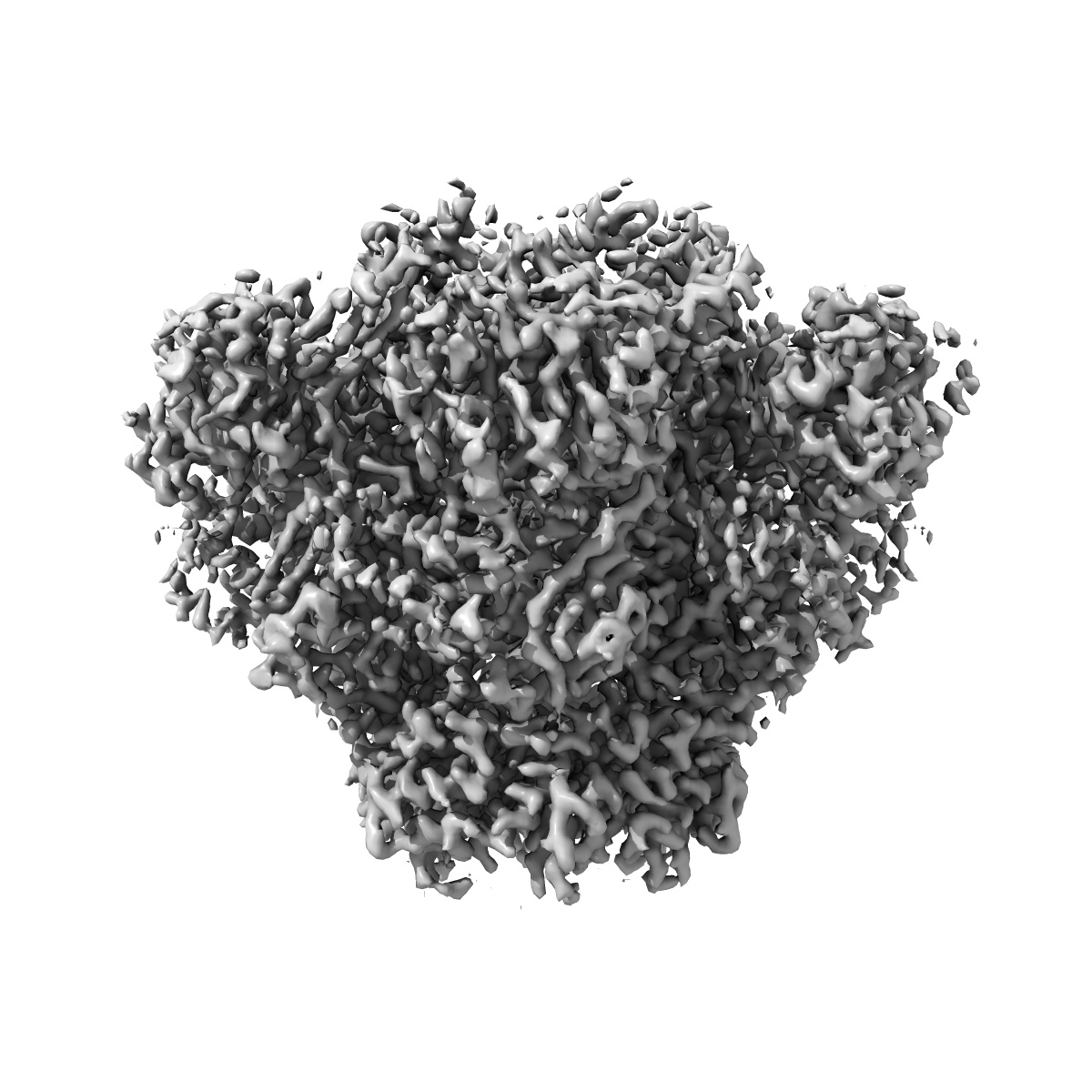
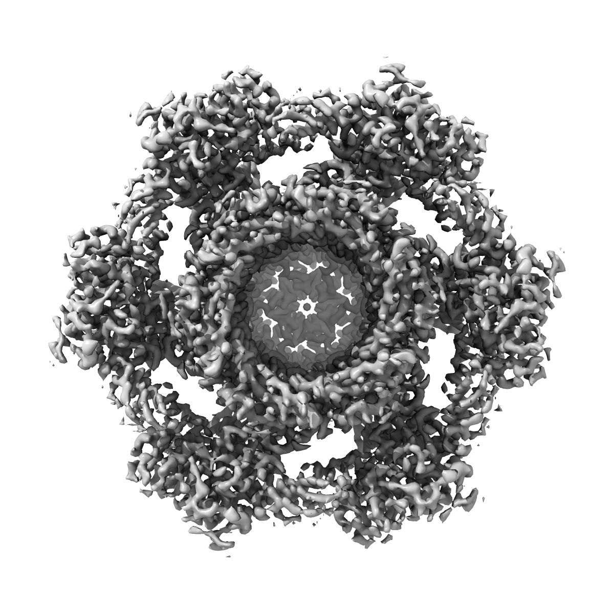
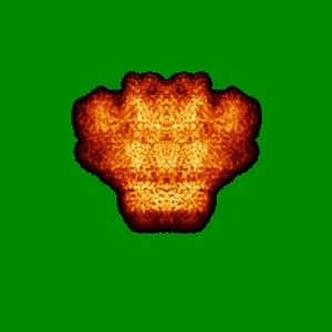
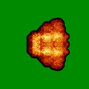
EMD-9763
Cryo-EM Structure of an Extracellular Contractile Injection System, PVC sheath/tube terminator in extended state
EMD-9763
Single-particle3.8 Å
 Deposition: 24/12/2018
Map released: 10/04/2019
Last modified: 27/03/2024
Buffer
pH: 7.4
Vitrification
Microscope: FEI TITAN KRIOS
Illumination mode: FLOOD BEAM
Imaging mode: BRIGHT FIELD
Electron source: FIELD EMISSION GUN
Acceleration voltage: 300 kV
Specimen holder model: FEI TITAN KRIOS AUTOGRID HOLDER
Cooling holder cryogen: NITROGEN
Alignment procedure: COMA FREE
Illumination mode: FLOOD BEAM
Imaging mode: BRIGHT FIELD
Electron source: FIELD EMISSION GUN
Acceleration voltage: 300 kV
Specimen holder model: FEI TITAN KRIOS AUTOGRID HOLDER
Cooling holder cryogen: NITROGEN
Alignment procedure: COMA FREE
Image Recording
[1]
Detector model:
FEI FALCON II (4k x 4k)
Detector mode: INTEGRATING
Average electron dose per image: 46.4 e/Å2
Detector mode: INTEGRATING
Average electron dose per image: 46.4 e/Å2
Final
reconstruction
Resolution: 3.8
Å
(
BY AUTHOR)
Resolution method: FSC 0.143 CUT-OFF
Number of images used: 45000
Resolution method: FSC 0.143 CUT-OFF
Number of images used: 45000
⌯ Applied Symmetry
Point group:
C6
Startup model
[1]
Type:
NONE
⦨ Initial angle
assignment
Type:
NOT APPLICABLE
⦩ Final angle assignment
Type:
PROJECTION MATCHING
Format: CCP4
Data type: IMAGE STORED AS FLOATING POINT NUMBER (4 BYTES)
Data type: IMAGE STORED AS FLOATING POINT NUMBER (4 BYTES)
⬡ Geometry
| X | Y | Z | |
|---|---|---|---|
| Origin | 0 | 0 | 0 |
| Dimensions (px) | 256 | 256 | 256 |
| Dimensions (Å) | 286.976 | 286.976 | 286.976 |
| Voxel size (Å) | 1.121 | 1.121 | 1.121 |
Contour list
| Primary | Level | Source |
|---|---|---|
| True | 0.14 | AUTHOR |