EMD-9524
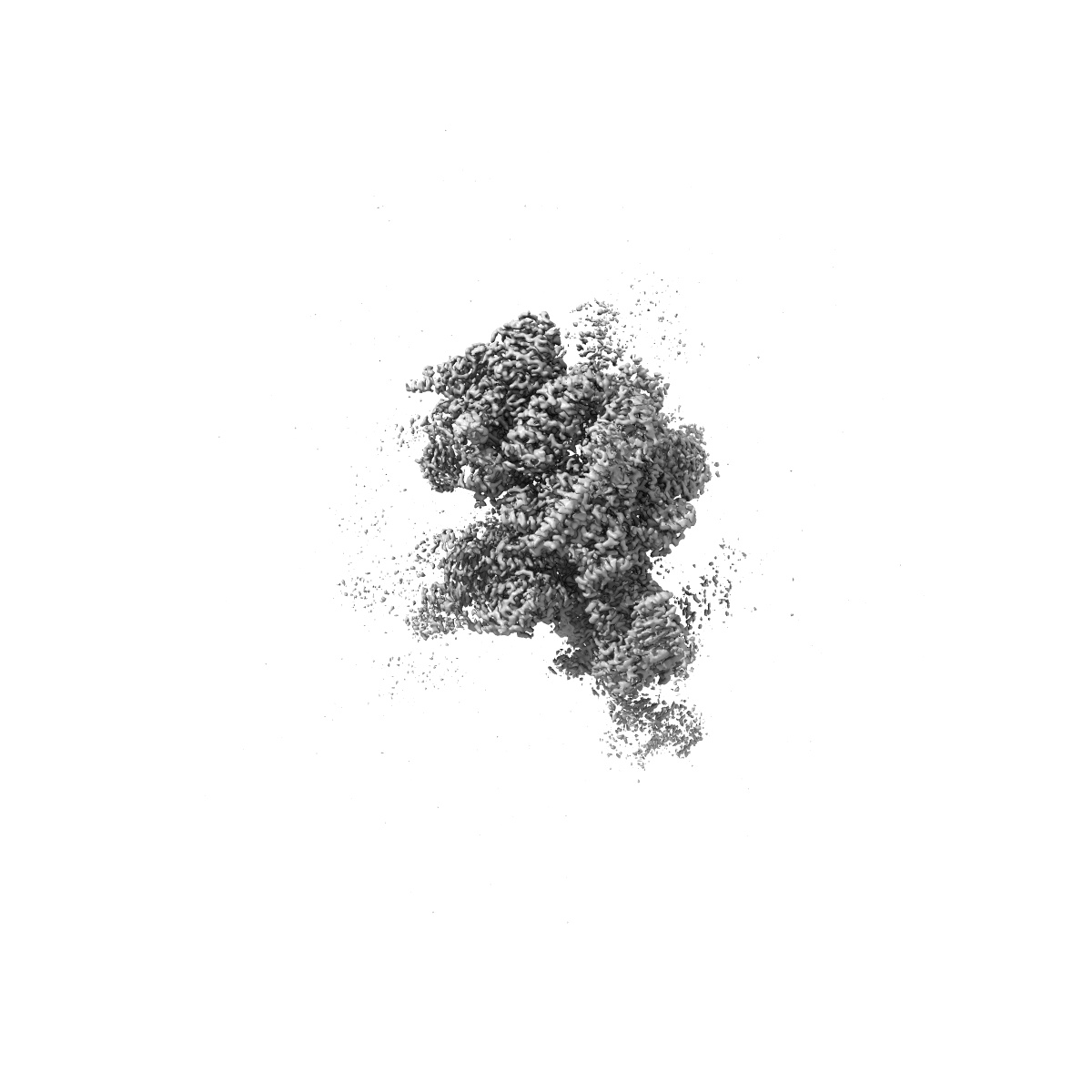
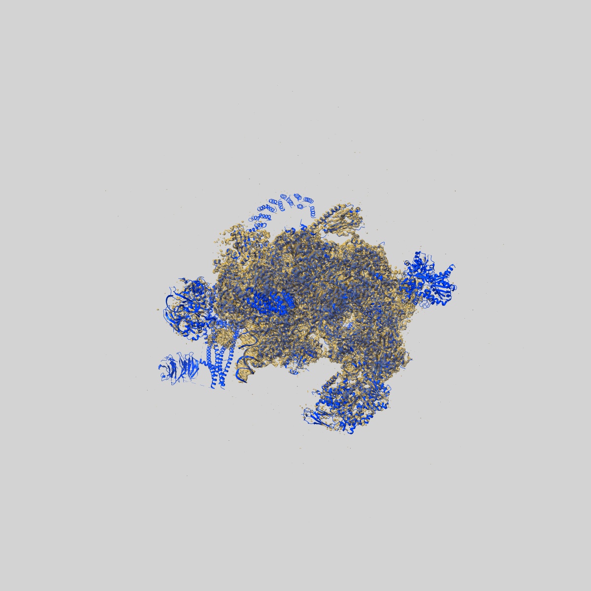
Cryo-EM structure of the activated spliceosome (Bact complex) at 3.5 angstrom resolution
EMD-9524
Single-particle3.5 Å
 Deposition: 18/08/2016
Map released: 21/09/2016
Last modified: 27/11/2024
Buffer
pH: 8.0
Details: CEB buffer (10 mM Tris-HCl, pH 8.0, 75 mM NaCl, 1 mM Mg(OAc)2, 1 mM imidazole, 0.01% NP40, 1 mM TCEP, 0.5 mM EGTA)
Details: CEB buffer (10 mM Tris-HCl, pH 8.0, 75 mM NaCl, 1 mM Mg(OAc)2, 1 mM imidazole, 0.01% NP40, 1 mM TCEP, 0.5 mM EGTA)
Vitrification
Cryogen name: ETHANE
Microscope: FEI TITAN KRIOS
Illumination mode: FLOOD BEAM
Imaging mode: BRIGHT FIELD
Electron source: FIELD EMISSION GUN
Acceleration voltage: 300 kV
Illumination mode: FLOOD BEAM
Imaging mode: BRIGHT FIELD
Electron source: FIELD EMISSION GUN
Acceleration voltage: 300 kV
Image Recording
[1]
Detector model:
GATAN K2 SUMMIT (4k x 4k)
Detector mode: SUPER-RESOLUTION
Scanner: OTHER
Average electron dose per image: 4.7 e/Å2
Detector mode: SUPER-RESOLUTION
Scanner: OTHER
Average electron dose per image: 4.7 e/Å2
Final
reconstruction
Startup model
[1]
Type:
EMDB MAP
⦨ Initial angle
assignment
Type:
ANGULAR RECONSTITUTION
⦩ Final angle assignment
Type:
ANGULAR RECONSTITUTION
Format: CCP4
Data type: IMAGE STORED AS FLOATING POINT NUMBER (4 BYTES)
Annotation details: Cryo-EM map of Bact spliceosome at 3.5-angstrom resolution
Data type: IMAGE STORED AS FLOATING POINT NUMBER (4 BYTES)
Annotation details: Cryo-EM map of Bact spliceosome at 3.5-angstrom resolution
⬡ Geometry
| X | Y | Z | |
|---|---|---|---|
| Origin | 0 | 0 | 0 |
| Dimensions (px) | 400 | 400 | 400 |
| Dimensions (Å) | 522.4 | 522.4 | 522.4 |
| Voxel size (Å) | 1.3060001 | 1.3060001 | 1.3060001 |
Contour list
| Primary | Level | Source |
|---|---|---|
| True | 0.0405 | AUTHOR |