EMD-8900
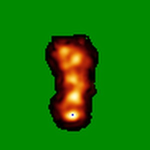
Negative stain reconstruction of human autophagy associated protein, ATG2A
EMD-8900
Single-particle29.4 Å
 Deposition: 21/08/2017
Map released: 06/09/2017
Last modified: 29/01/2020
Concentration: 0.01
mg/mL
Buffer
pH: 7.5
Staining
Grid
Mesh: 400
Model: Maxtaform
Material: COPPER/RHODIUM
Model: Maxtaform
Material: COPPER/RHODIUM
Pretreatment
Support Film [1]
| Material | Topology | Thickness |
|---|---|---|
| CARBON | CONTINUOUS | - |
Microscope: FEI TECNAI SPIRIT
Illumination mode: FLOOD BEAM
Imaging mode: BRIGHT FIELD
Electron source: LAB6
Acceleration voltage: 120 kV
C2 aperture diameter: 100.0 µm
Nominal CS: 2.2 mm
Nominal defocus: 1.0 µm - 1.5 µm
Calibrated defocus: 0.62 µm - 2.0 µm
Nominal magnification: 52000.0
Calibrated magnification: 52000.0
Specimen holder model: SIDE ENTRY, EUCENTRIC
Alignment procedure: BASIC
Temperature
Minimum: 273.15
K
Maximum: 298.15 K
Maximum: 298.15 K
Image Recording
[1]
Detector model:
TVIPS TEMCAM-F416 (4k x 4k)
Dimensions: 4096 pixel x 4096 pixel
Sampling interval: 15.6 µm
Number of grids: 1
Number of real images: 590
Average exposure time: 0.42 s
Average electron dose per image: 20.0 e/Å2
Dimensions: 4096 pixel x 4096 pixel
Sampling interval: 15.6 µm
Number of grids: 1
Number of real images: 590
Average exposure time: 0.42 s
Average electron dose per image: 20.0 e/Å2
Final
reconstruction
Resolution: 29.4
Å
(
BY AUTHOR)
Resolution method: FSC 0.143 CUT-OFF
Number of images used: 6386
Resolution method: FSC 0.143 CUT-OFF
Number of images used: 6386
⌯ Applied Symmetry
Point group:
C1
Software
[1]
| Name | Version | Details |
|---|---|---|
| RELION | 1.4 | - |
Startup model
[1]
⦨ Initial angle
assignment
Type:
PROJECTION MATCHING
⦩ Final angle assignment
Particle selection
[1]
| Selected | Ref. model | Method | Software | Details |
|---|---|---|---|---|
| 37021 | - | - | - | - |
Final 3D classification
Software
[1]
| Name | Version | Details |
|---|---|---|
| RELION | 1.4 | - |
CTF correction
Software
[1]
| Name | Version | Details |
|---|---|---|
| RELION | 1.4 | - |
Format: CCP4
Data type: IMAGE STORED AS FLOATING POINT NUMBER (4 BYTES)
Annotation details: Negative stain reconstruction of human ATG2A protein
Data type: IMAGE STORED AS FLOATING POINT NUMBER (4 BYTES)
Annotation details: Negative stain reconstruction of human ATG2A protein
⬡ Geometry
| X | Y | Z | |
|---|---|---|---|
| Origin | 0 | 0 | 0 |
| Dimensions (px) | 96 | 96 | 96 |
| Dimensions (Å) | 393.59998 | 393.59998 | 393.59998 |
| Voxel size (Å) | 4.1 | 4.1 | 4.1 |
Contour list
| Primary | Level | Source |
|---|---|---|
| True | 5.2 | AUTHOR |