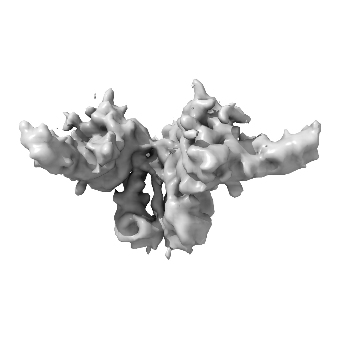
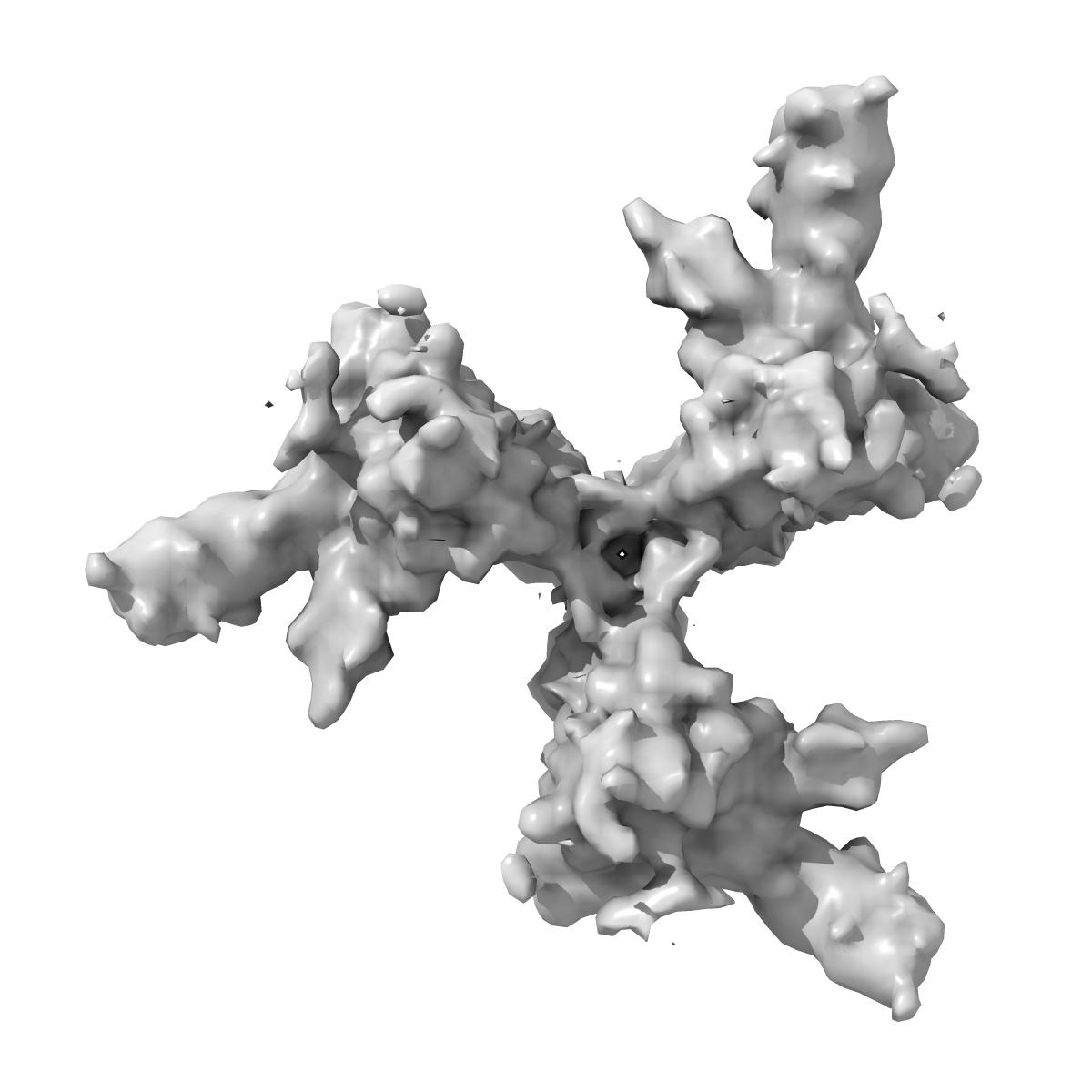
EMD-8729
Cryo-EM reconstruction of the HIV-1 BG505 SOSIP.664 Env trimer in complex with soluble CD4 (D1-D2)
EMD-8729
Single-particle10.5 Å
 Deposition: 13/05/2017
Map released: 26/07/2017
Last modified: 29/01/2020
Concentration: 1
mg/mL
Buffer
pH: 7.4
Buffer components [3]:
Buffer components [3]:
| Name | Formula | Concentration | ChEBI |
|---|---|---|---|
| Sodium chloride | NaCl | 150.0 mM | |
| Tris | C4H11NO3 | 50.0 mM | |
| DDM | - | 1.125 mM | - |
Grid
Vitrification
Cryogen name: ETHANE
Chamber humidity: 50%
Chamber temperature: 277 K
Instrument: HOMEMADE PLUNGER
Details: 5 microliters of the complex was incubated with 3 microliters of a fresh 1.8 mM DDM solution. A 3 microliter aliquot of the complex was applied to a C-Flat grid (CF-2/2-4C, Electron Microscopy Sciences, Protochips, Inc.) which had been plasma cleaned for 5 seconds using a mixture of Ar/O2 (Gatan Solarus 950 Plasma system), blotted off, and then immediately plunged into liquid ethane using a manual freeze plunger..
Microscope: FEI TITAN KRIOS
Illumination mode: FLOOD BEAM
Imaging mode: BRIGHT FIELD
Electron source: FIELD EMISSION GUN
Acceleration voltage: 300 kV
C2 aperture diameter: 100.0 µm
Nominal CS: 2.7 mm
Calibrated defocus: 1.0 µm - 4.0 µm
Nominal magnification: 22500.0
Calibrated magnification: 38167.0
Specimen holder model: FEI TITAN KRIOS AUTOGRID HOLDER
Cooling holder cryogen: NITROGEN
Alignment procedure: COMA FREE
Temperature
Minimum: 90.0
K
Maximum: 90.0 K
Maximum: 90.0 K
Image Recording
[1]
Detector model:
GATAN K2 SUMMIT (4k x 4k)
Detector mode: COUNTING
Dimensions: 3838 pixel x 3710 pixel
Sampling interval: 5.0 µm
Frames per image: 1-28
Number of grids: 1
Average exposure time: 5.6 s
Average electron dose per image: 32.0 e/Å2
Details: Individual frames were gain-corrected, aligned, and summed using MotionCor.
Detector mode: COUNTING
Dimensions: 3838 pixel x 3710 pixel
Sampling interval: 5.0 µm
Frames per image: 1-28
Number of grids: 1
Average exposure time: 5.6 s
Average electron dose per image: 32.0 e/Å2
Details: Individual frames were gain-corrected, aligned, and summed using MotionCor.
Final
reconstruction
Resolution: 10.5
Å
(
BY AUTHOR)
Resolution method: FSC 0.143 CUT-OFF
Number of images used: 1754
Algorithm: FOURIER SPACE
Resolution method: FSC 0.143 CUT-OFF
Number of images used: 1754
Algorithm: FOURIER SPACE
⌯ Applied Symmetry
Point group:
C3
Software
[1]
| Name | Version | Details |
|---|---|---|
| FREALIGN | 9.11 | - |
Startup model
[1]
Type:
INSILICO MODEL
Insilico model: Common lines model using OptiMod
Details:An initial model was generated directly from the class averages using OptiMod.
Insilico model: Common lines model using OptiMod
Details:An initial model was generated directly from the class averages using OptiMod.
⦨ Initial angle
assignment
Type:
PROJECTION MATCHING
Angular sampling: 7.5 degrees
Details: Relion 3D classification, auto mode
Angular sampling: 7.5 degrees
Details: Relion 3D classification, auto mode
Software
[1]
| Name | Version | Details |
|---|---|---|
| RELION | 1.3 | - |
⦩ Final angle assignment
Type:
PROJECTION MATCHING
Details: Frealign 3D classification and refinement
Details: Frealign 3D classification and refinement
Software
[1]
| Name | Version | Details |
|---|---|---|
| FREALIGN | 9.11 | - |
Final 3D classification
Software
[1]
| Name | Version | Details |
|---|---|---|
| FREALIGN | 3.11 | - |
CTF correction
Details: Performed internally in Relion and Frealign
Software
[1]
| Name | Version | Details |
|---|---|---|
| CTFFIND | 3 | - |
Format: CCP4
Data type: IMAGE STORED AS FLOATING POINT NUMBER (4 BYTES)
Annotation details: BG505 SOSIP trimer bound to sCD4
Data type: IMAGE STORED AS FLOATING POINT NUMBER (4 BYTES)
Annotation details: BG505 SOSIP trimer bound to sCD4
⬡ Geometry
| X | Y | Z | |
|---|---|---|---|
| Origin | 0 | 0 | 0 |
| Dimensions (px) | 128 | 128 | 128 |
| Dimensions (Å) | 335.36 | 335.36 | 335.36 |
| Voxel size (Å) | 2.62 | 2.62 | 2.62 |
Contour list
| Primary | Level | Source |
|---|---|---|
| True | 0.11 | AUTHOR |