






EMD-8681
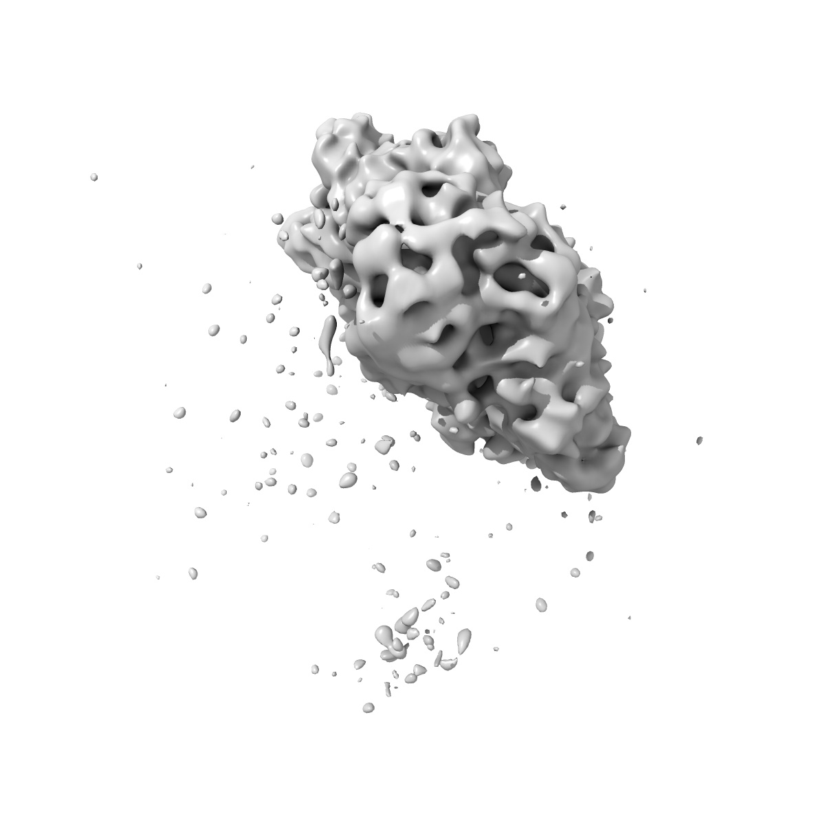
Conformational Landscape of the p28-Bound Human Proteasome Regulatory Particle
EMD-8681
Single-particle7.9 Å
 Deposition: 13/04/2017
Map released: 23/08/2017
Last modified: 13/03/2024
Buffer
pH: 7.5
Vitrification
Cryogen name: ETHANE
Microscope: FEI TECNAI ARCTICA
Illumination mode: FLOOD BEAM
Imaging mode: BRIGHT FIELD
Electron source: FIELD EMISSION GUN
Acceleration voltage: 200 kV
Illumination mode: FLOOD BEAM
Imaging mode: BRIGHT FIELD
Electron source: FIELD EMISSION GUN
Acceleration voltage: 200 kV
Image Recording
[1]
Final
reconstruction
Startup model
[1]
Type:
INSILICO MODEL
⦨ Initial angle
assignment
Type:
ANGULAR RECONSTITUTION
⦩ Final angle assignment
Type:
PROJECTION MATCHING
Format: CCP4
Data type: IMAGE STORED AS FLOATING POINT NUMBER (4 BYTES)
Annotation details: Final Map of Rpn1-p28-AAA subcomplex in the TA5 state, corrected with a B-factor of -450
Data type: IMAGE STORED AS FLOATING POINT NUMBER (4 BYTES)
Annotation details: Final Map of Rpn1-p28-AAA subcomplex in the TA5 state, corrected with a B-factor of -450
⬡ Geometry
| X | Y | Z | |
|---|---|---|---|
| Origin | 0 | 0 | 0 |
| Dimensions (px) | 360 | 360 | 360 |
| Dimensions (Å) | 309.6 | 309.6 | 309.6 |
| Voxel size (Å) | 0.86 | 0.86 | 0.86 |
Contour list
| Primary | Level | Source |
|---|---|---|
| True | 0.004 | AUTHOR |