EMD-8620
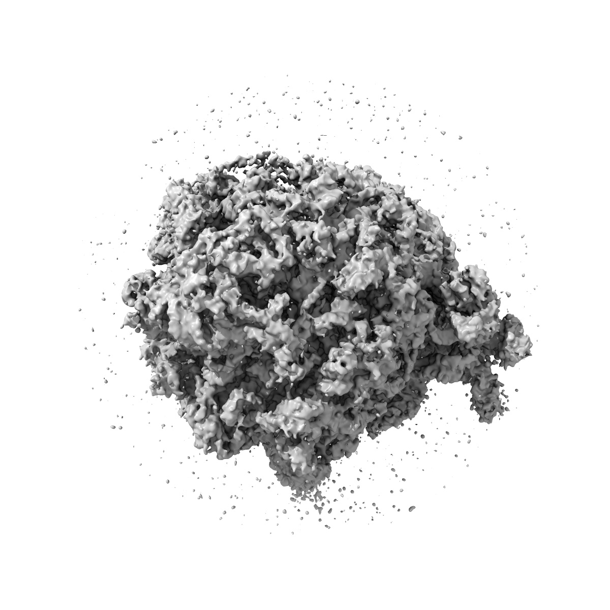
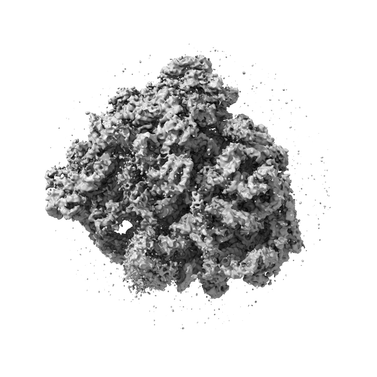
70S ribosome bound with near-cognate ternary complex base-paired to A site codon, closed 30S (Structure III-nc)
EMD-8620
Single-particle3.8 Å
 Deposition: 24/02/2017
Map released: 07/06/2017
Last modified: 13/03/2024
Details: 125 nM 50S, 125 nM 30S, 625 nM mRNA, 250 nM fMet-tRNAfMet, 1.25 micromolar EF-Tu, 500 micromolar GDPCP, 1.25 micromolar Lys-tRNALys
Buffer
pH: 7.5
Buffer components [6]:
Buffer components [6]:
| Name | Formula | Concentration | ChEBI |
|---|---|---|---|
| HEPES | - | 20.0 mM | - |
| Magnesium chloride | MgCl2 | 20.0 mM | |
| Ammonium chloride | NH4Cl | 150.0 mM | |
| Spermidine | - | 2.0 mM | - |
| Spermine | - | 0.1 mM | - |
| Beta-mercaptoethanol | - | 6.0 mM | - |
Grid
Mesh: 400
Model: C-flat-1.2/1.3
Material: COPPER
Model: C-flat-1.2/1.3
Material: COPPER
Pretreatment
Support Film [1]
| Material | Topology | Thickness |
|---|---|---|
| CARBON | HOLEY ARRAY | - |
Vitrification
Cryogen name: ETHANE
Chamber humidity: 90%
Chamber temperature: 275 K
Instrument: GATAN CRYOPLUNGE 3
Details: 2 uL of complex was applied to each grid. After a 10-second incubation, the grids were blotted for 2 to 4 seconds..
Chamber humidity: 90%
Chamber temperature: 275 K
Instrument: GATAN CRYOPLUNGE 3
Details: 2 uL of complex was applied to each grid. After a 10-second incubation, the grids were blotted for 2 to 4 seconds..
Microscope: FEI TITAN KRIOS
Illumination mode: FLOOD BEAM
Imaging mode: BRIGHT FIELD
Electron source: FIELD EMISSION GUN
Acceleration voltage: 300 kV
Nominal CS: 2.7 mm
Nominal defocus: 0.5 µm - 5.0 µm
Nominal magnification: 60976.0
Calibrated magnification: 60976.0
Specimen holder model: FEI TITAN KRIOS AUTOGRID HOLDER
Cooling holder cryogen: NITROGEN
Alignment procedure: COMA FREE
Image Recording
[1]
Detector model:
GATAN K2 SUMMIT (4k x 4k)
Detector mode: SUPER-RESOLUTION
Dimensions: 7676 pixel x 7420 pixel
Frames per image: 1-30
Number of grids: 3
Number of real images: 1773
Average exposure time: 0.4 s
Average electron dose per image: 1.0 e/Å2
Detector mode: SUPER-RESOLUTION
Dimensions: 7676 pixel x 7420 pixel
Frames per image: 1-30
Number of grids: 3
Number of real images: 1773
Average exposure time: 0.4 s
Average electron dose per image: 1.0 e/Å2
Details: Gain reference was applied, movies were aligned, and the summed imaged were corrected for magnification anisotropy.
Final
reconstruction
Resolution: 3.8
Å
(
BY AUTHOR)
Resolution method: FSC 0.143 CUT-OFF
Number of classed used: 3
Number of images used: 5758
Algorithm: BACK PROJECTION
Details:
Resolution method: FSC 0.143 CUT-OFF
Number of classed used: 3
Number of images used: 5758
Algorithm: BACK PROJECTION
Details:
⌯ Applied Symmetry
Point group:
C1
Software
[1]
| Name | Version | Details |
|---|---|---|
| FREALIGN | 9.11 | - |
⦨ Initial angle
assignment
⦩ Final angle assignment
Particle selection
[1]
| Selected | Ref. model | Method | Software | Details |
|---|---|---|---|---|
| 572417 | - | - | - | Particles were picked from micrographs using Signature reference-based particle picker. |
Final 3D classification
Number of classes:
6
Avg. number of members per classes: 50000.0
Avg. number of members per classes: 50000.0
Software
[1]
| Name | Version | Details |
|---|---|---|
| FREALIGN | 9.11 | - |
Format: CCP4
Data type: IMAGE STORED AS FLOATING POINT NUMBER (4 BYTES)
Annotation details: 70S ribosome bound with near-cognate ternary complex base-paired to A site codon, closed 30S (Structure III-nc): filtered map from Frealign (FFILT setting in Frealign = T), used for model building after B-factor sharpening. This volume not B-factor sharpened.
⬡ Geometry
| X | Y | Z | |
|---|---|---|---|
| Origin | 0 | 0 | 0 |
| Dimensions (px) | 480 | 480 | 480 |
| Dimensions (Å) | 393.6 | 393.6 | 393.6 |
| Voxel size (Å) | 0.82 | 0.82 | 0.82 |
Contour list
| Primary | Level | Source |
|---|---|---|
| True | 2.85 | AUTHOR |