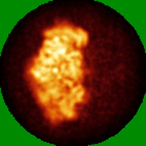
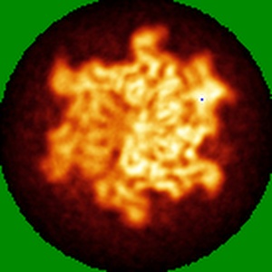
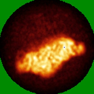
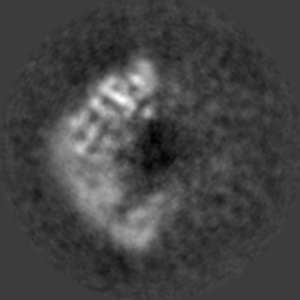
EMD-8553
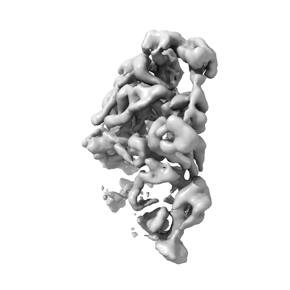
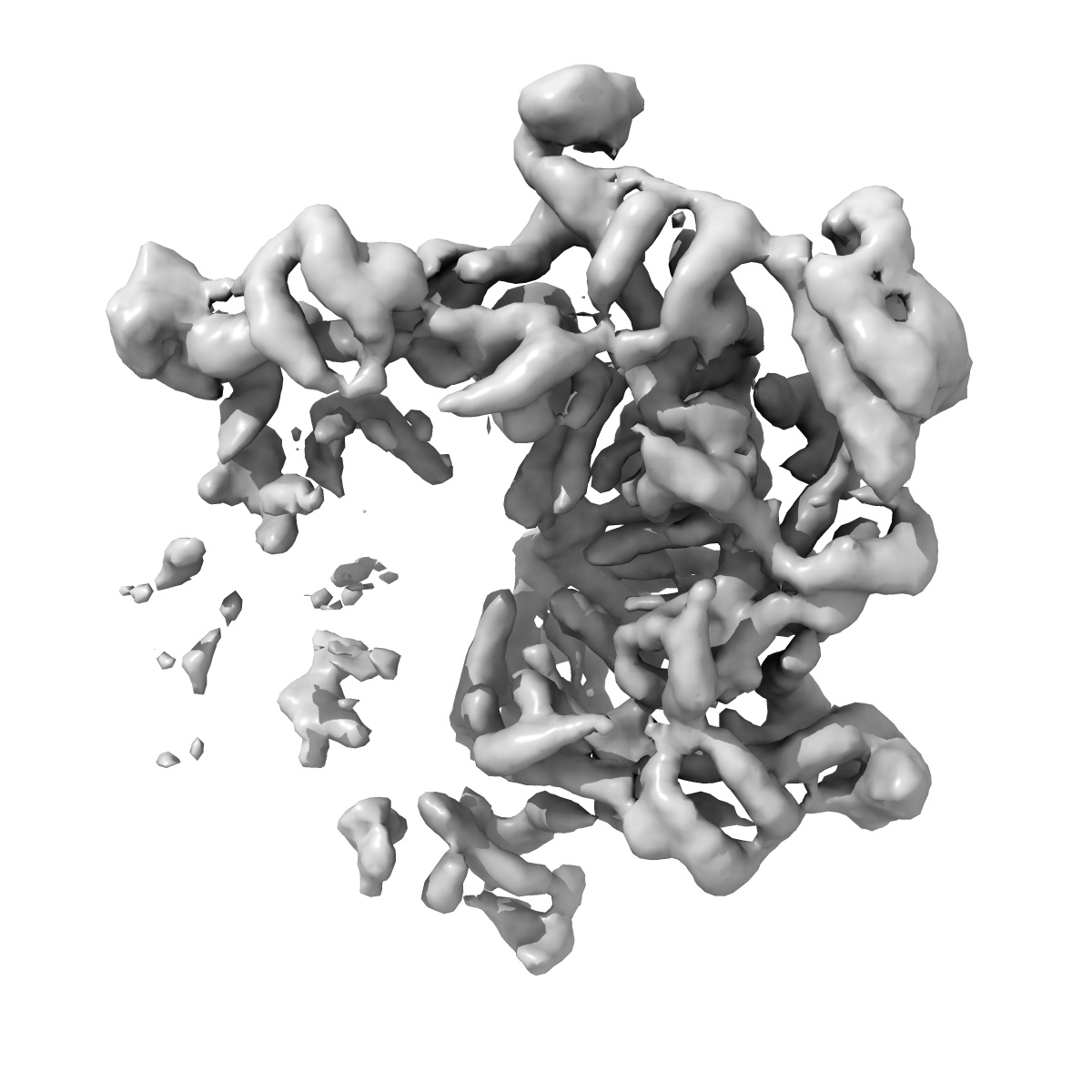
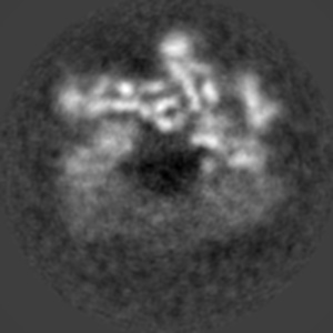
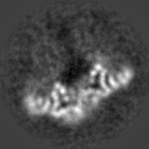
Vps4-Vta1 complex, VSL_B
EMD-8553
Single-particle6.7 Å
 Deposition: 13/01/2017
Map released: 12/04/2017
Last modified: 29/01/2020
Buffer
pH: 7.4
Grid
Material:
COPPER
Vitrification
Cryogen name: ETHANE
Chamber humidity: 80%
Chamber temperature: 277 K
Instrument: FEI VITROBOT MARK III
Chamber humidity: 80%
Chamber temperature: 277 K
Instrument: FEI VITROBOT MARK III
Microscope: FEI TECNAI F20
Illumination mode: FLOOD BEAM
Imaging mode: BRIGHT FIELD
Electron source: FIELD EMISSION GUN
Acceleration voltage: 200 kV
Illumination mode: FLOOD BEAM
Imaging mode: BRIGHT FIELD
Electron source: FIELD EMISSION GUN
Acceleration voltage: 200 kV
Image Recording
[1]
Detector model:
GATAN K2 SUMMIT (4k x 4k)
Detector mode: COUNTING
Average exposure time: 0.2 s
Average electron dose per image: 1.3 e/Å2
Detector mode: COUNTING
Average exposure time: 0.2 s
Average electron dose per image: 1.3 e/Å2
Final
reconstruction
Resolution: 6.7
Å
(
BY AUTHOR)
Resolution method: FSC 0.143 CUT-OFF
Number of images used: 38421
Resolution method: FSC 0.143 CUT-OFF
Number of images used: 38421
Software
[1]
| Name | Version | Details |
|---|---|---|
| RELION | 1.4 | - |
Startup model
[1]
⦨ Initial angle
assignment
Type:
ANGULAR RECONSTITUTION
⦩ Final angle assignment
Type:
ANGULAR RECONSTITUTION
Format: CCP4
Data type: IMAGE STORED AS FLOATING POINT NUMBER (4 BYTES)
Annotation details: Vps4-Vta1 complex, VSL_B
Data type: IMAGE STORED AS FLOATING POINT NUMBER (4 BYTES)
Annotation details: Vps4-Vta1 complex, VSL_B
⬡ Geometry
| X | Y | Z | |
|---|---|---|---|
| Origin | 0 | 0 | 0 |
| Dimensions (px) | 138 | 138 | 138 |
| Dimensions (Å) | 194.442 | 194.442 | 194.442 |
| Voxel size (Å) | 1.409 | 1.409 | 1.409 |
Contour list
| Primary | Level | Source |
|---|---|---|
| True | 0.0253 | AUTHOR |