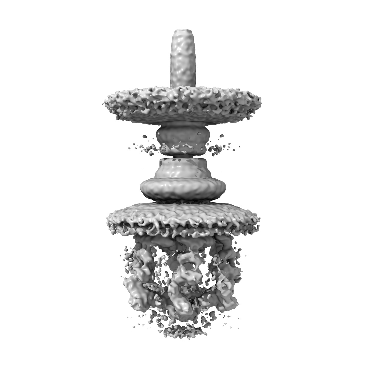
EMD-8544
Salmonella Type III secretion injectisome
EMD-8544
Subtomogram averaging17.0 Å
 Deposition: 04/01/2017
Map released: 29/03/2017
Last modified: 29/01/2020
Buffer
pH: 7.0
Vitrification
Cryogen name: ETHANE
Microscope: FEI POLARA 300
Illumination mode: OTHER
Imaging mode: DARK FIELD
Electron source: FIELD EMISSION GUN
Acceleration voltage: 300 kV
Illumination mode: OTHER
Imaging mode: DARK FIELD
Electron source: FIELD EMISSION GUN
Acceleration voltage: 300 kV
Image Recording
[1]
Detector model:
GATAN K2 SUMMIT (4k x 4k)
Detector mode: COUNTING
Average electron dose per image: 1.4 e/Å2
Detector mode: COUNTING
Average electron dose per image: 1.4 e/Å2
Final
reconstruction
Resolution: 17.0
Å
(
BY AUTHOR)
Resolution method: FSC 0.5 CUT-OFF
Resolution method: FSC 0.5 CUT-OFF
⌯ Applied Symmetry
Point group:
C6
⦩ Final angle assignment
Extraction
Number of images used: 5274
Software
[1]
| Name | Version | Details |
|---|---|---|
| IMOD | - | - |
Format: CCP4
Data type: IMAGE STORED AS FLOATING POINT NUMBER (4 BYTES)
Annotation details: Salmonella injectisome
Data type: IMAGE STORED AS FLOATING POINT NUMBER (4 BYTES)
Annotation details: Salmonella injectisome
⬡ Geometry
| X | Y | Z | |
|---|---|---|---|
| Origin | -200 | -200 | -200 |
| Dimensions (px) | 400 | 400 | 400 |
| Dimensions (Å) | 1040.0 | 1040.0 | 1040.0 |
| Voxel size (Å) | 2.6 | 2.6 | 2.6 |
Contour list
| Primary | Level | Source |
|---|---|---|
| True | 2.53 | AUTHOR |