EMD-8539

Single-particle
4.6 Å
EMD-8539 Deposition: 22/12/2016
Map released: 10/05/2017
Last modified: 25/12/2024
Overview 3D View Sample Experiment Validation Additional data Links

EMD-8539

Archive Files (Depositor)

Primary 3D volume (map.gz) Half-map 1 Half-map 2 (map.gz) Experimental metadata (xml) Experimental metadata (cif.gz)

Validation (wwPDB)

Map-only validation report (pdf.gz) Map-5ucy summary report (pdf.gz) Map-5ucy FULL report (pdf.gz)

EMDB Annotations

EMICSS entry mapping (xml)

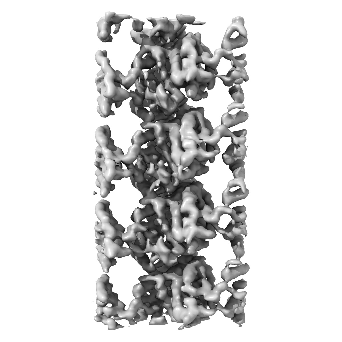
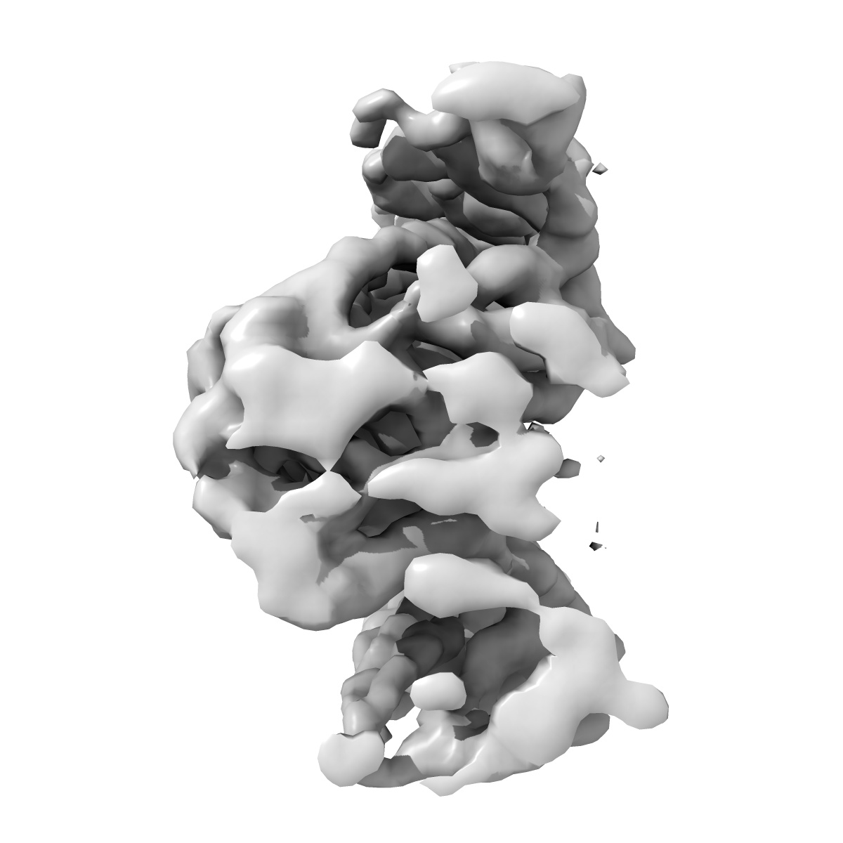
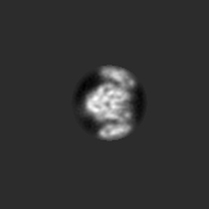
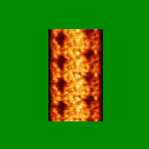
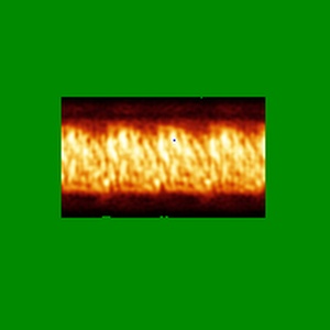
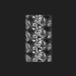
Cryo-EM map of protofilament of microtubule doublet

EMD-8539

Single-particle
4.6 Å
EMD-8539 Deposition: 22/12/2016
Map released: 10/05/2017
Last modified: 25/12/2024
Overview 3D View Sample Experiment Validation Additional data Links
Sample Organism: Tetrahymena thermophila
Sample: Protofilament from ciliary microtubule doublet
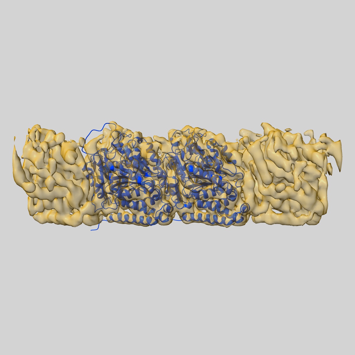
Fitted models: 5ucy

Deposition Authors: Ichikawa M , Liu D
Subnanometre-resolution structure of the doublet microtubule reveals new classes of microtubule-associated proteins.
Ichikawa M , Liu D, Kastritis PL, Basu K, Hsu TC, Yang S, Bui KH
(2017) Nat Commun , 8 , 15035 - 15035
PUBMED: 28462916
DOI: doi:10.1038/ncomms15035
ISSN: 2041-1723