EMD-8243
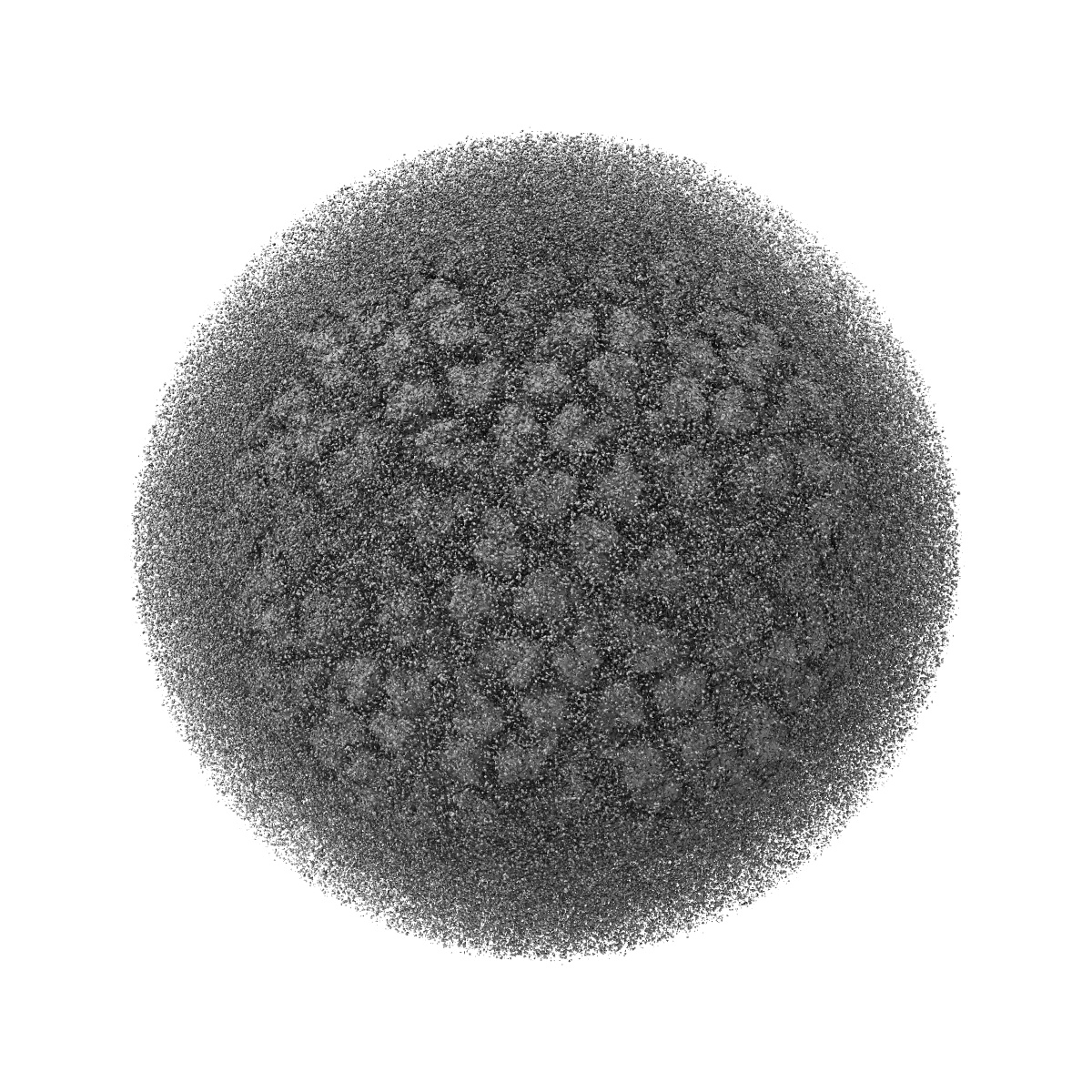
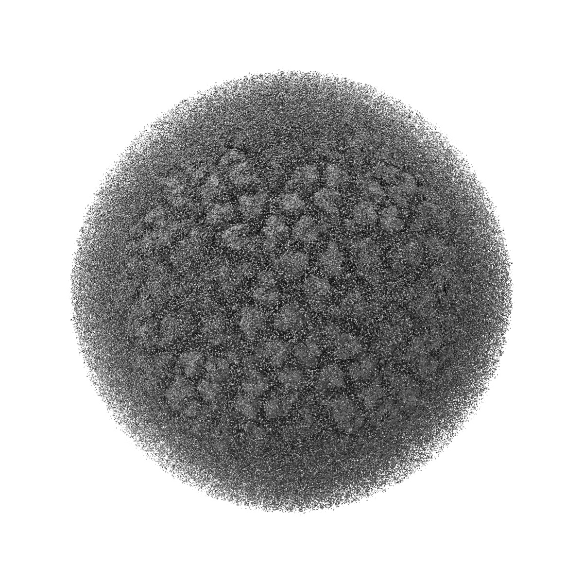
Near-atomic resolution cryo-EM reconstruction of HPV16 complexed with Fab V5
EMD-8243
Single-particle4.7 Å
 Deposition: 10/06/2016
Map released: 13/12/2017
Last modified: 24/10/2018
Buffer
pH: 7.4
Vitrification
Cryogen name: ETHANE
Microscope: FEI POLARA 300
Illumination mode: SPOT SCAN
Imaging mode: BRIGHT FIELD
Electron source: FIELD EMISSION GUN
Acceleration voltage: 300 kV
Illumination mode: SPOT SCAN
Imaging mode: BRIGHT FIELD
Electron source: FIELD EMISSION GUN
Acceleration voltage: 300 kV
Image Recording
[1]
Final
reconstruction
Startup model
[1]
⦨ Initial angle
assignment
Type:
COMMON LINE
⦩ Final angle assignment
Type:
COMMON LINE
Format: CCP4
Data type: IMAGE STORED AS FLOATING POINT NUMBER (4 BYTES)
Annotation details: HPV16 complexed with Fab V5
Data type: IMAGE STORED AS FLOATING POINT NUMBER (4 BYTES)
Annotation details: HPV16 complexed with Fab V5
⬡ Geometry
| X | Y | Z | |
|---|---|---|---|
| Origin | -260 | -260 | -260 |
| Dimensions (px) | 520 | 520 | 520 |
| Dimensions (Å) | 910.0 | 910.0 | 910.0 |
| Voxel size (Å) | 1.75 | 1.75 | 1.75 |
Contour list
| Primary | Level | Source |
|---|---|---|
| True | 1.0 | AUTHOR |