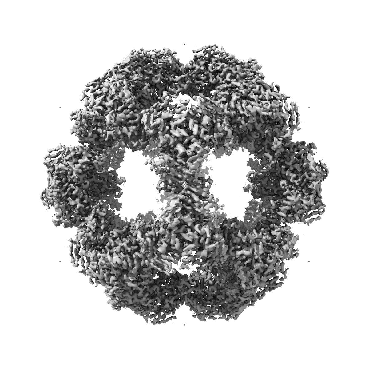
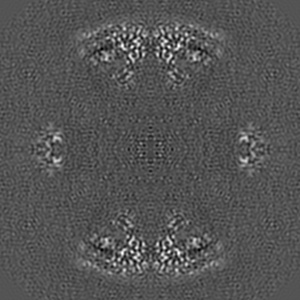
EMD-7610
Atomic Structure of the E2 Inner Core of Human Pyruvate Dehydrogenase Complex
EMD-7610
Single-particle3.1 Å
 Deposition: 21/03/2018
Map released: 18/04/2018
Last modified: 13/03/2024
Buffer
pH: 7.2
Vitrification
Cryogen name: ETHANE
Microscope: FEI TITAN KRIOS
Illumination mode: FLOOD BEAM
Imaging mode: BRIGHT FIELD
Electron source: FIELD EMISSION GUN
Acceleration voltage: 300 kV
Illumination mode: FLOOD BEAM
Imaging mode: BRIGHT FIELD
Electron source: FIELD EMISSION GUN
Acceleration voltage: 300 kV
Image Recording
[1]
Detector model:
GATAN K2 SUMMIT (4k x 4k)
Detector mode: COUNTING
Average exposure time: 8.0 s
Average electron dose per image: 38.0 e/Å2
Detector mode: COUNTING
Average exposure time: 8.0 s
Average electron dose per image: 38.0 e/Å2
Final
reconstruction
Resolution: 3.1
Å
(
BY AUTHOR)
Resolution method: FSC 0.143 CUT-OFF
Number of images used: 49374
Resolution method: FSC 0.143 CUT-OFF
Number of images used: 49374
Software
[1]
| Name | Version | Details |
|---|---|---|
| RELION | - | - |
Startup model
[1]
Type:
NONE
⦨ Initial angle
assignment
⦩ Final angle assignment
Format: CCP4
Data type: IMAGE STORED AS FLOATING POINT NUMBER (4 BYTES)
Annotation details: 3D reconstruction of PDC tE2 after particle polishing
Data type: IMAGE STORED AS FLOATING POINT NUMBER (4 BYTES)
Annotation details: 3D reconstruction of PDC tE2 after particle polishing
⬡ Geometry
| X | Y | Z | |
|---|---|---|---|
| Origin | -128 | -128 | -128 |
| Dimensions (px) | 256 | 256 | 256 |
| Dimensions (Å) | 261.12 | 261.12 | 261.12 |
| Voxel size (Å) | 1.02 | 1.02 | 1.02 |
Contour list
| Primary | Level | Source |
|---|---|---|
| True | 2.5 | AUTHOR |