





EMD-7493
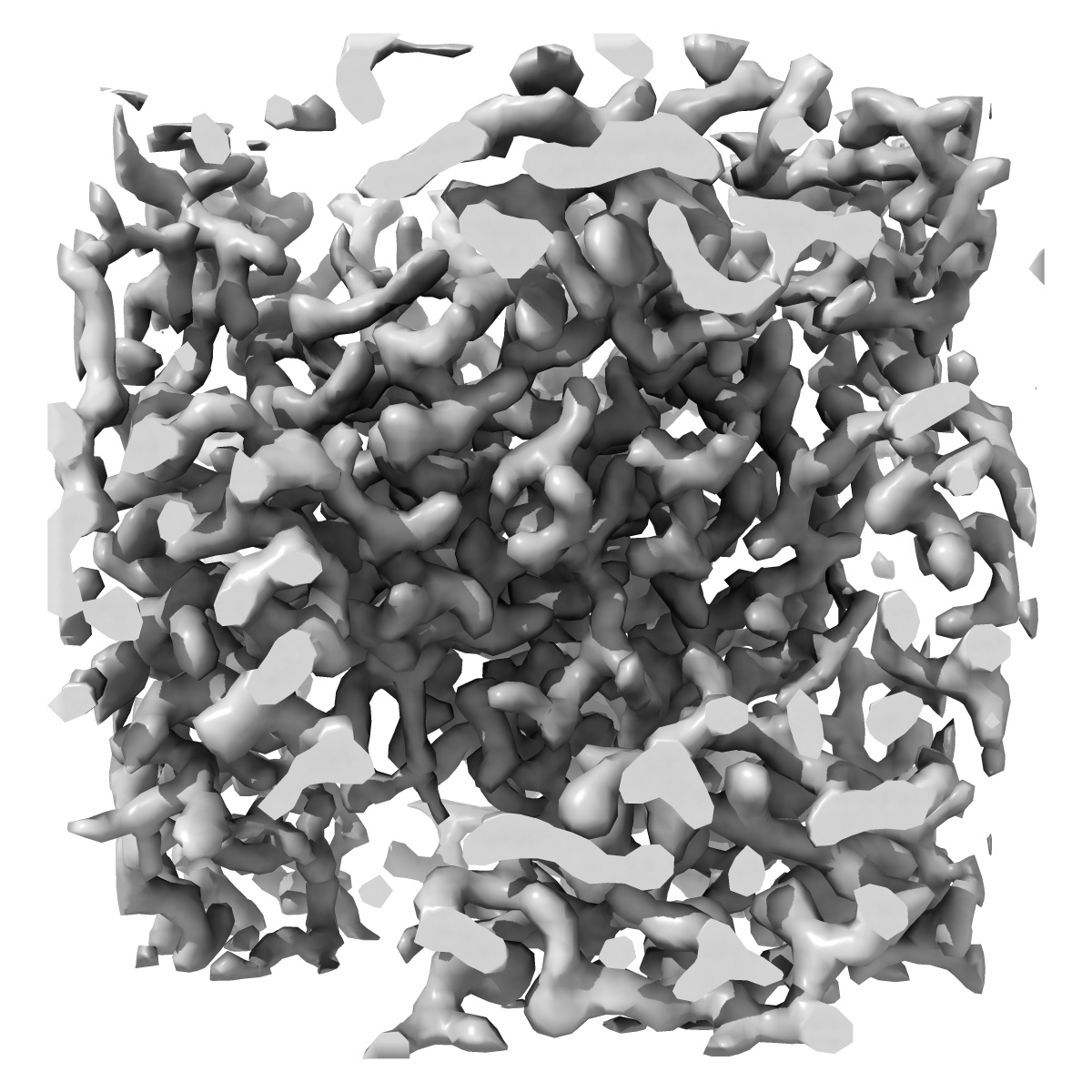
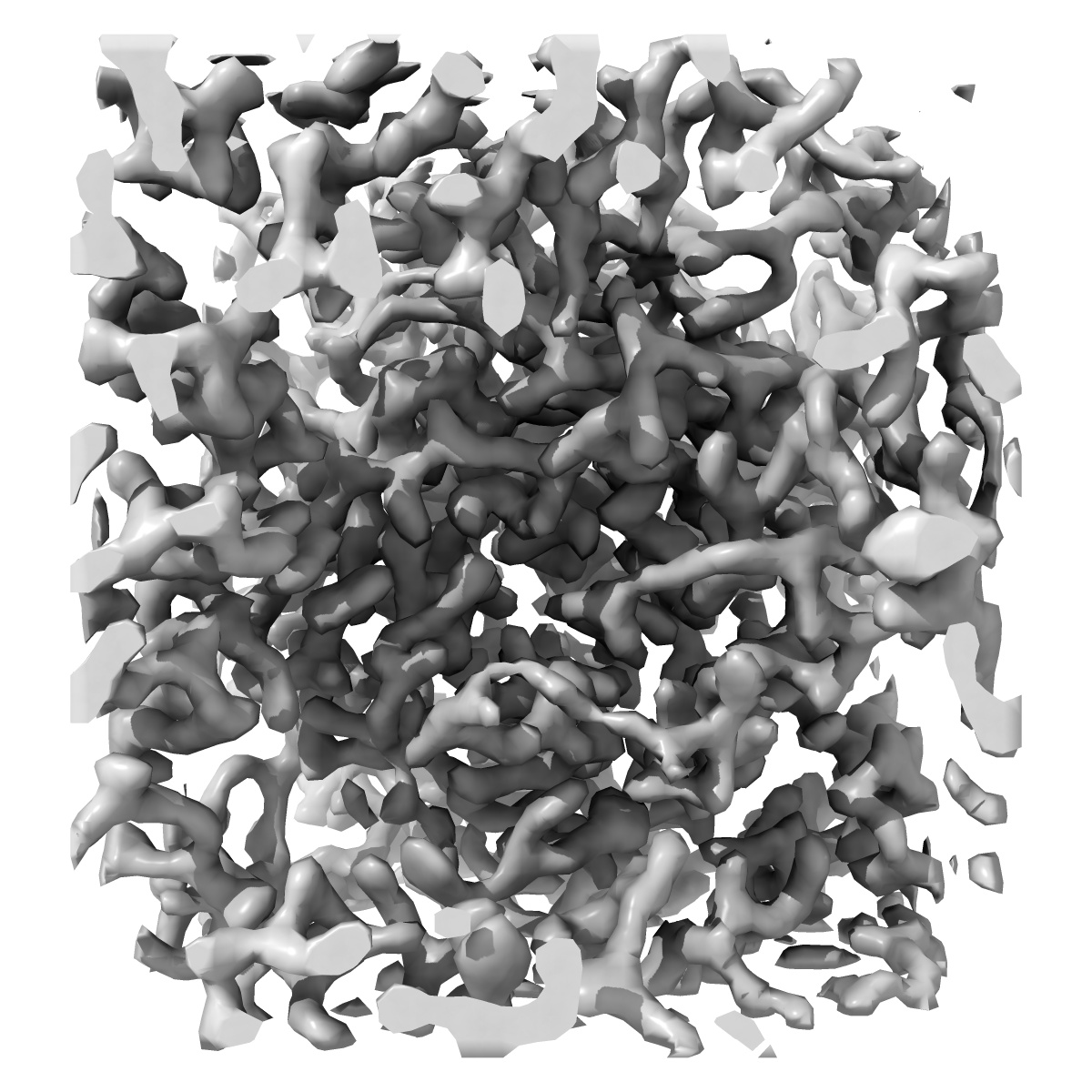
2.80 A MicroED structure of proteinase K at 6.0 e- / A^2
EMD-7493
Electron Crystallography2.8 Å
 Deposition: 02/03/2018
Map released: 16/05/2018
Last modified: 16/10/2024
Concentration: 25
mg/mL
Buffer
pH: 8.0
Buffer components [2]:
Buffer components [2]:
| Name | Formula | Concentration | ChEBI |
|---|---|---|---|
| Ammonium sulfate | N2H8SO4 | 1.2 M | |
| Tris | C4H11NO3 | 0.1 M |
Vitrification
Microscope: FEI TECNAI F20
Illumination mode: FLOOD BEAM
Imaging mode: DIFFRACTION
Electron source: FIELD EMISSION GUN
Acceleration voltage: 200 kV
Camera Length: 1200 mm
Cooling holder cryogen: NITROGEN
Illumination mode: FLOOD BEAM
Imaging mode: DIFFRACTION
Electron source: FIELD EMISSION GUN
Acceleration voltage: 200 kV
Camera Length: 1200 mm
Cooling holder cryogen: NITROGEN
Image Recording
[1]
Detector model:
TVIPS TEMCAM-F416 (4k x 4k)
Dimensions: 2048 pixel x 2048 pixel
Number of grids: 1
Number of real images: 240
Number of diffraction images: 240
Average exposure time: 5.1 s
Average electron dose per image: 0.0357 e/Å2
Dimensions: 2048 pixel x 2048 pixel
Number of grids: 1
Number of real images: 240
Number of diffraction images: 240
Average exposure time: 5.1 s
Average electron dose per image: 0.0357 e/Å2
Final
reconstruction
Molecular replacement
Software
[1]
| Name | Version | Details |
|---|---|---|
| MOLREP | 11.4.05 | - |
⌯ Symmetry determination
Software [1]
| Name | Version | Details |
|---|---|---|
| POINTLESS | 1.11.3 | - |
Merging
Software [1]
| Name | Version | Details |
|---|---|---|
| AIMLESS | 0.5.32 | - |
Crystallography statistics
Number of intensities measured:
30596
Number of structure factors: 5423
Fourier space coverage: 88.4
R sym: 0.536
R merge: 0.536
Overall phase error: 62.59
Overall phase residual: 62.59
Phase error rejection criteria: 0
High resolution: 2.8 Å
Number of structure factors: 5423
Fourier space coverage: 88.4
R sym: 0.536
R merge: 0.536
Overall phase error: 62.59
Overall phase residual: 62.59
Phase error rejection criteria: 0
High resolution: 2.8 Å
Shell list
[1]
| High resolution | Low resolution | Number of structure factors | Phase residual | Fourier space coverage | Multiplicity |
|---|---|---|---|---|---|
| 2.8 Å | 2.87 Å | 374 | 71.28 | 88.21 | 5.8 |
Format: CCP4
Data type: IMAGE STORED AS FLOATING POINT NUMBER (4 BYTES)
Annotation details: 2.80 A MicroED density map of proteinase K at 6.0 e- / A^2
Data type: IMAGE STORED AS FLOATING POINT NUMBER (4 BYTES)
Annotation details: 2.80 A MicroED density map of proteinase K at 6.0 e- / A^2
⬡ Geometry
| X | Y | Z | |
|---|---|---|---|
| Origin | -47 | -5 | -31 |
| Dimensions (px) | 58 | 54 | 62 |
| Dimensions (Å) | 67.4502 | 67.4502 | 100.8179 |
| Voxel size (Å) | 0.93680835 | 0.93680835 | 0.90015984 |
Contour list
| Primary | Level | Source |
|---|---|---|
| True | 0.19626 | AUTHOR |