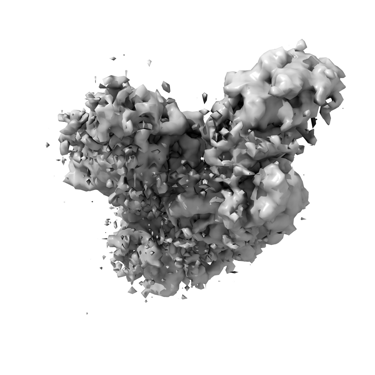
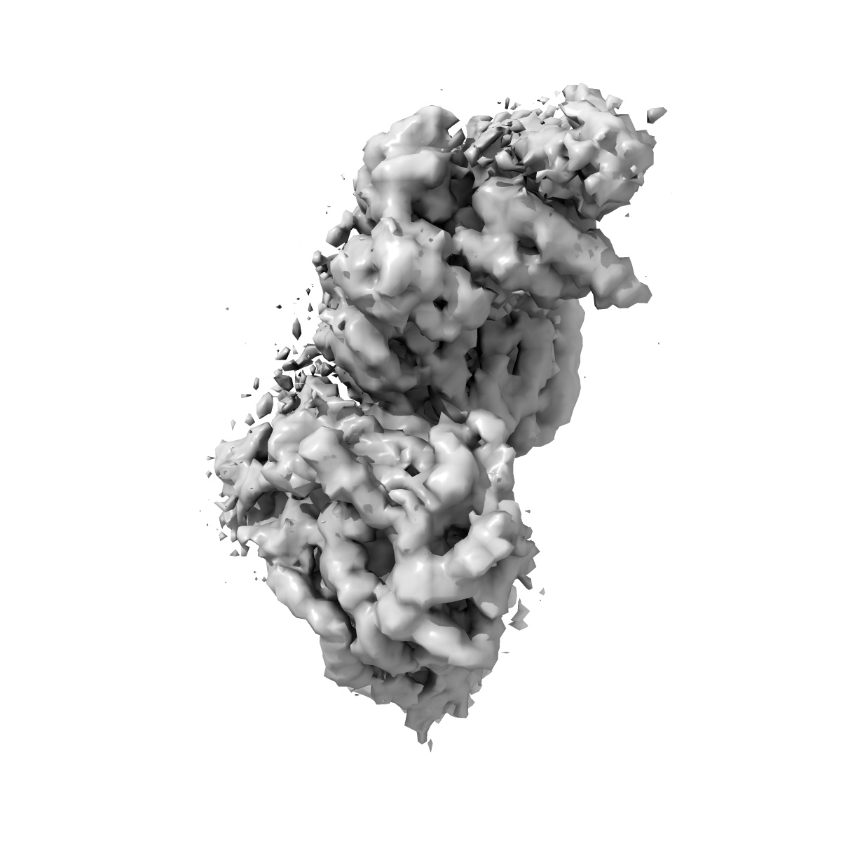
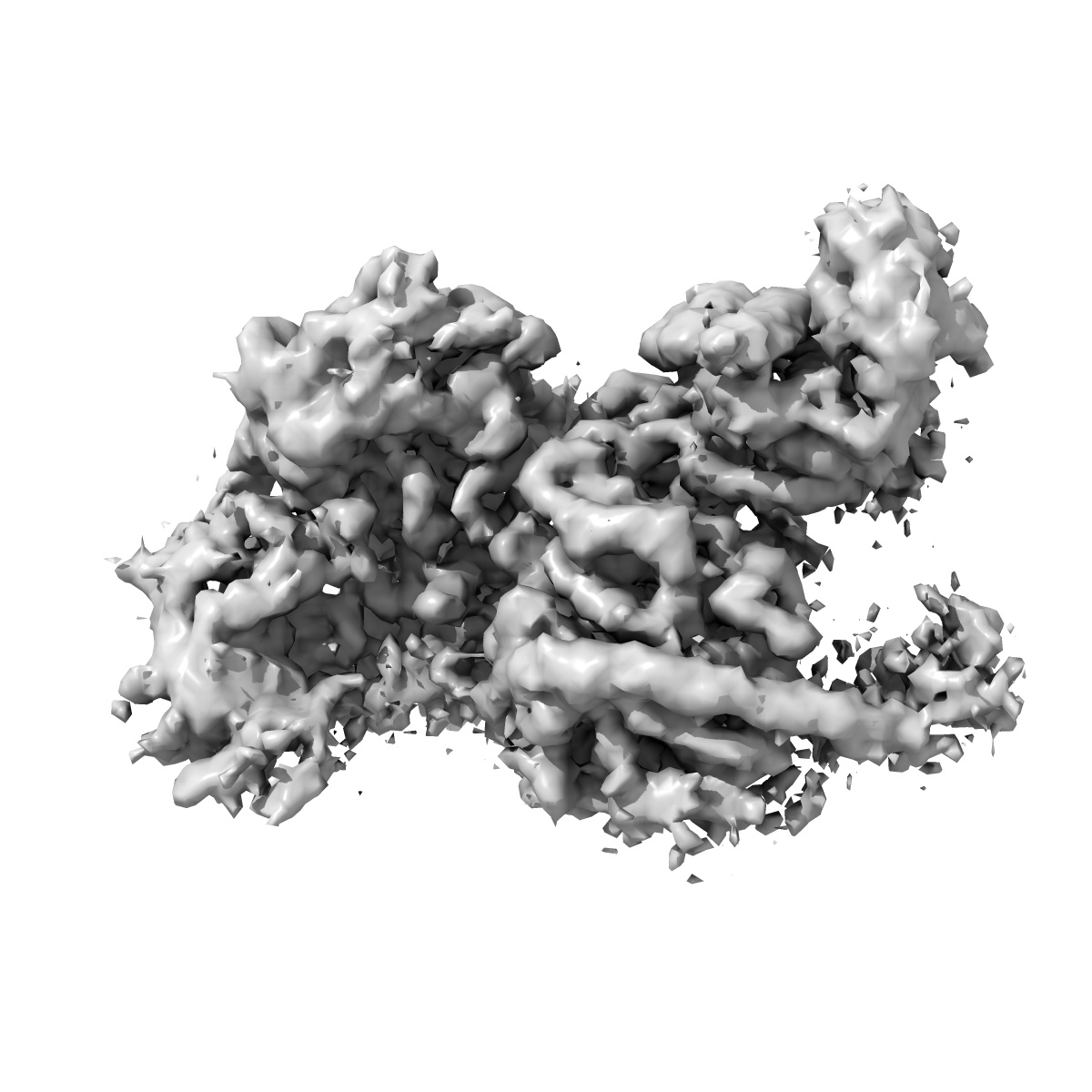
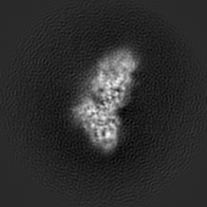
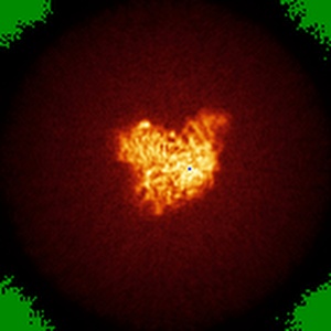
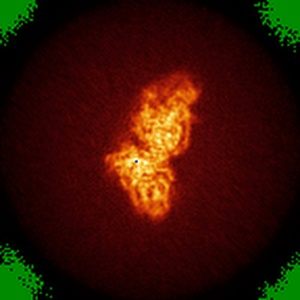
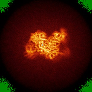
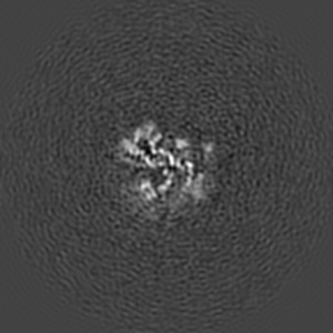
EMD-7337
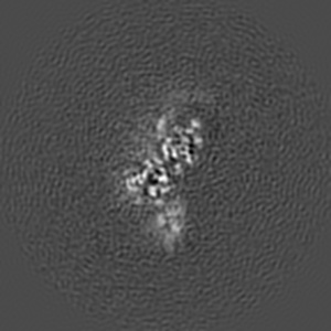
Cryo-EM structure of PRC2 bound to cofactors AEBP2 and JARID2 in the Extended Basal state
EMD-7337
Single-particle4.6 Å
 Deposition: 08/01/2018
Map released: 31/01/2018
Last modified: 11/12/2019
Buffer
pH: 7.9
Vitrification
Cryogen name: ETHANE
Microscope: FEI TITAN KRIOS
Illumination mode: SPOT SCAN
Imaging mode: BRIGHT FIELD
Electron source: FIELD EMISSION GUN
Acceleration voltage: 300 kV
Illumination mode: SPOT SCAN
Imaging mode: BRIGHT FIELD
Electron source: FIELD EMISSION GUN
Acceleration voltage: 300 kV
Image Recording
[1]
Format: CCP4
Data type: IMAGE STORED AS FLOATING POINT NUMBER (4 BYTES)
Annotation details: Sharpened map (EB state)
Data type: IMAGE STORED AS FLOATING POINT NUMBER (4 BYTES)
Annotation details: Sharpened map (EB state)
⬡ Geometry
| X | Y | Z | |
|---|---|---|---|
| Origin | 0 | 0 | 0 |
| Dimensions (px) | 144 | 144 | 144 |
| Dimensions (Å) | 299.52 | 299.52 | 299.52 |
| Voxel size (Å) | 2.08 | 2.08 | 2.08 |
Contour list
| Primary | Level | Source |
|---|---|---|
| True | 0.08 | AUTHOR |