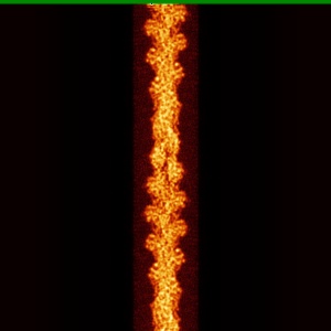
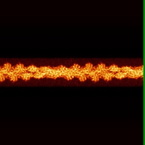
EMD-7115
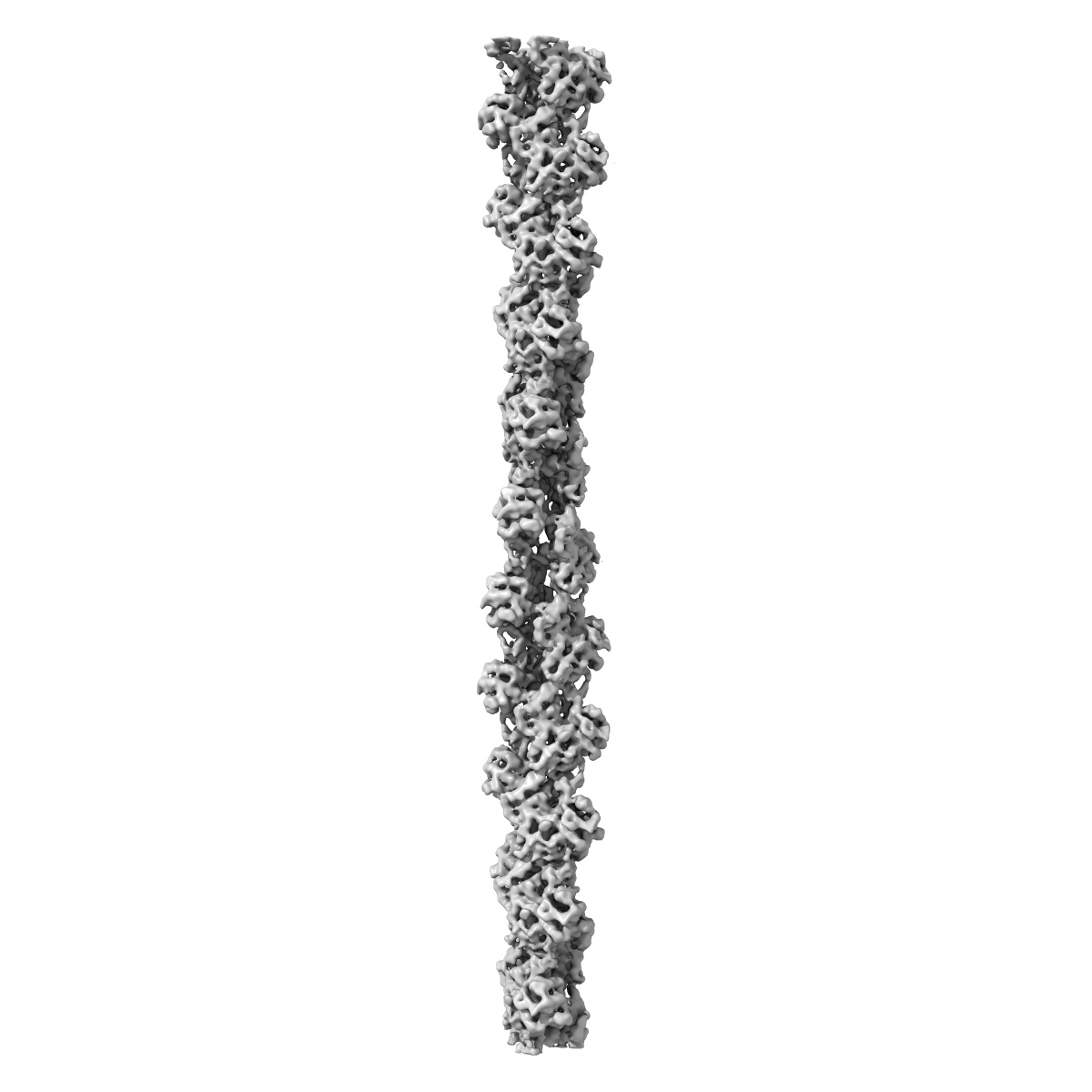
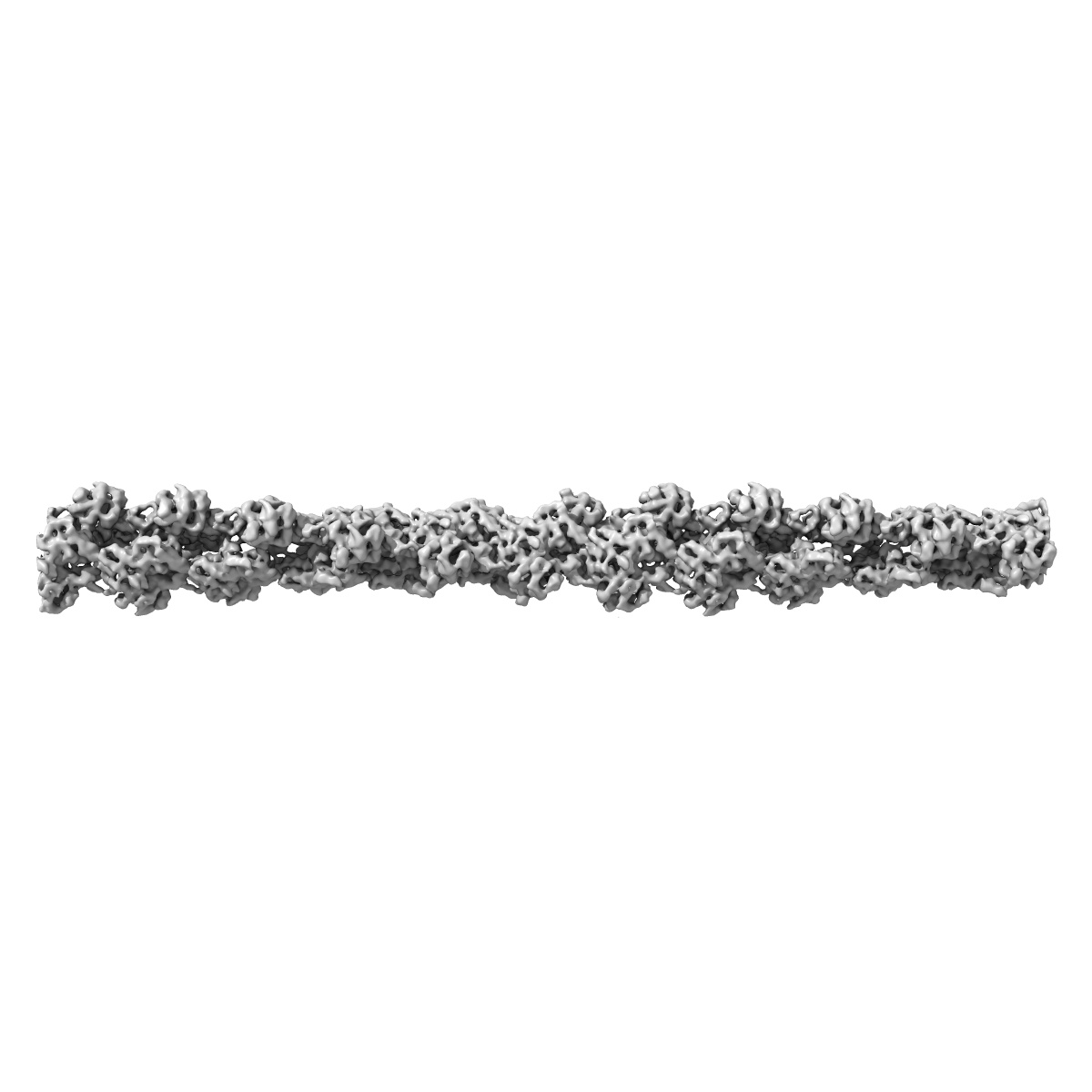
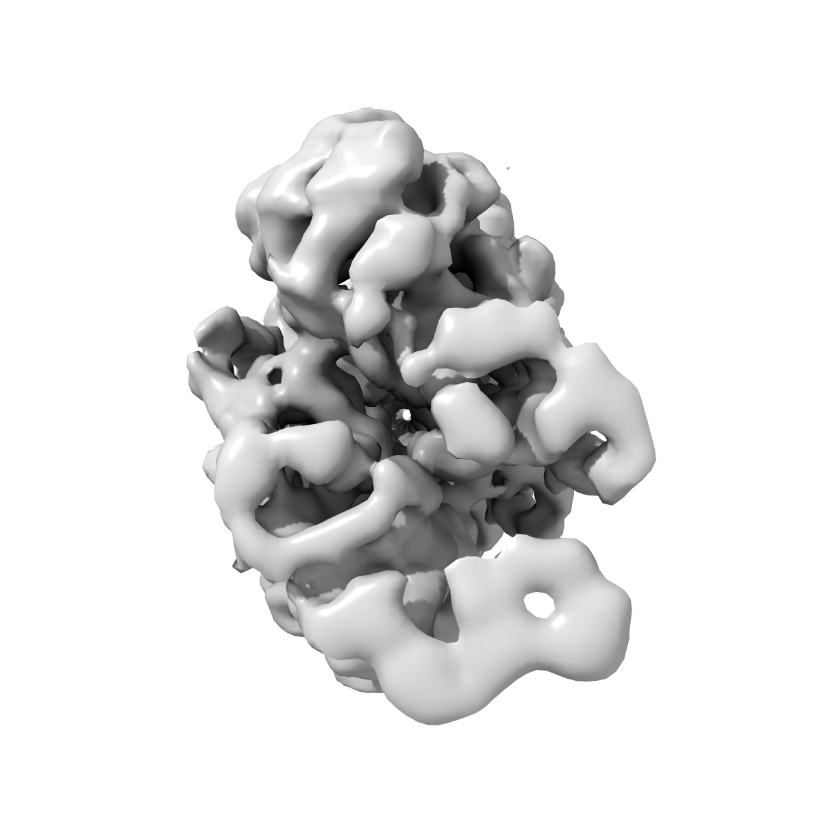
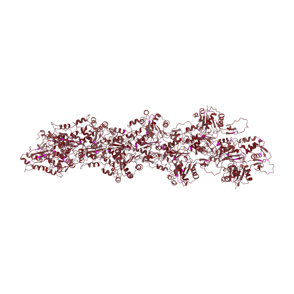
Structure of bare actin filament
EMD-7115
Helical reconstruction5.5 Å
 Deposition: 17/11/2017
Map released: 10/01/2018
Last modified: 13/03/2024
Concentration: 0.025
mg/mL
Details: Filamentous bare actin
Details: Filamentous bare actin
Buffer
pH: 7.5
Buffer components [8]:
Details: Buffer was filtered through 0.44 um filter and degassed.
Buffer components [8]:
| Name | Formula | Concentration | ChEBI |
|---|---|---|---|
| Potassium chloride | KCl | 50.0 mM | |
| Magnesium chloride | MgCl2 | 1.0 mM | |
| EGTA | C14H24N2O10 | 1.0 mM | |
| Imidazole | C3H4N2 | 10.0 mM | |
| Tris hydrochloride | C4H11NO3 | 2.0 mM | |
| Dithiothreitol | C4H10O2S2 | 0.5 mM | |
| Adenosine triphosphate | C10H16N5O13P3 | 200.0 mM | |
| Sodium azide | NaN3 | 0.01 % |
Grid
Vitrification
Cryogen name: ETHANE
Chamber humidity: 95%
Chamber temperature: 298 K
Instrument: LEICA EM GP
Details: Sample was applied to a glow-discharged holey carbon grid, incubated for 60 seconds and blotted for 3 seconds from the backside with filter paper..
Chamber humidity: 95%
Chamber temperature: 298 K
Instrument: LEICA EM GP
Details: Sample was applied to a glow-discharged holey carbon grid, incubated for 60 seconds and blotted for 3 seconds from the backside with filter paper..
Microscope: FEI TECNAI 20
Illumination mode: FLOOD BEAM
Imaging mode: BRIGHT FIELD
Electron source: FIELD EMISSION GUN
Acceleration voltage: 200 kV
C2 aperture diameter: 100.0 µm
Nominal CS: 2.0 mm
Nominal defocus: 1.5 µm - 3.0 µm
Nominal magnification: 29000.0
Specimen holder model: GATAN 626 SINGLE TILT LIQUID NITROGEN CRYO TRANSFER HOLDER
Cooling holder cryogen: NITROGEN
Alignment procedure: COMA FREE
Image Recording
[1]
Detector model:
GATAN K2 SUMMIT (4k x 4k)
Detector mode: COUNTING
Dimensions: 3838 pixel x 3710 pixel
Frames per image: 1-24
Number of grids: 1
Number of real images: 442
Average exposure time: 0.25 s
Average electron dose per image: 1.5 e/Å2
Detector mode: COUNTING
Dimensions: 3838 pixel x 3710 pixel
Frames per image: 1-24
Number of grids: 1
Number of real images: 442
Average exposure time: 0.25 s
Average electron dose per image: 1.5 e/Å2
Final
reconstruction
Resolution: 5.5
Å
(
BY AUTHOR)
Resolution method: FSC 0.143 CUT-OFF
Number of classed used: 1
Number of images used: 63139
Algorithm: FOURIER SPACE
Resolution method: FSC 0.143 CUT-OFF
Number of classed used: 1
Number of images used: 63139
Algorithm: FOURIER SPACE
⌯ Applied Symmetry
Software
[1]
| Name | Version | Details |
|---|---|---|
| FREALIGN | 9.11 | - |
⦩ Final angle assignment
Format: CCP4
Data type: IMAGE STORED AS FLOATING POINT NUMBER (4 BYTES)
Annotation details: CryoEM density map of actin alone, sharpened with a -350 B factor
Data type: IMAGE STORED AS FLOATING POINT NUMBER (4 BYTES)
Annotation details: CryoEM density map of actin alone, sharpened with a -350 B factor
⬡ Geometry
| X | Y | Z | |
|---|---|---|---|
| Origin | -256 | -256 | -256 |
| Dimensions (px) | 512 | 512 | 512 |
| Dimensions (Å) | 650.24 | 650.24 | 650.24 |
| Voxel size (Å) | 1.27 | 1.27 | 1.27 |
Contour list
| Primary | Level | Source |
|---|---|---|
| True | 12.0 | AUTHOR |