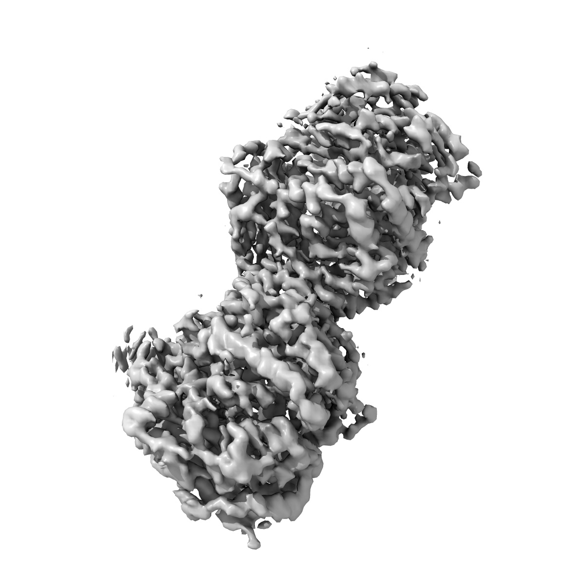
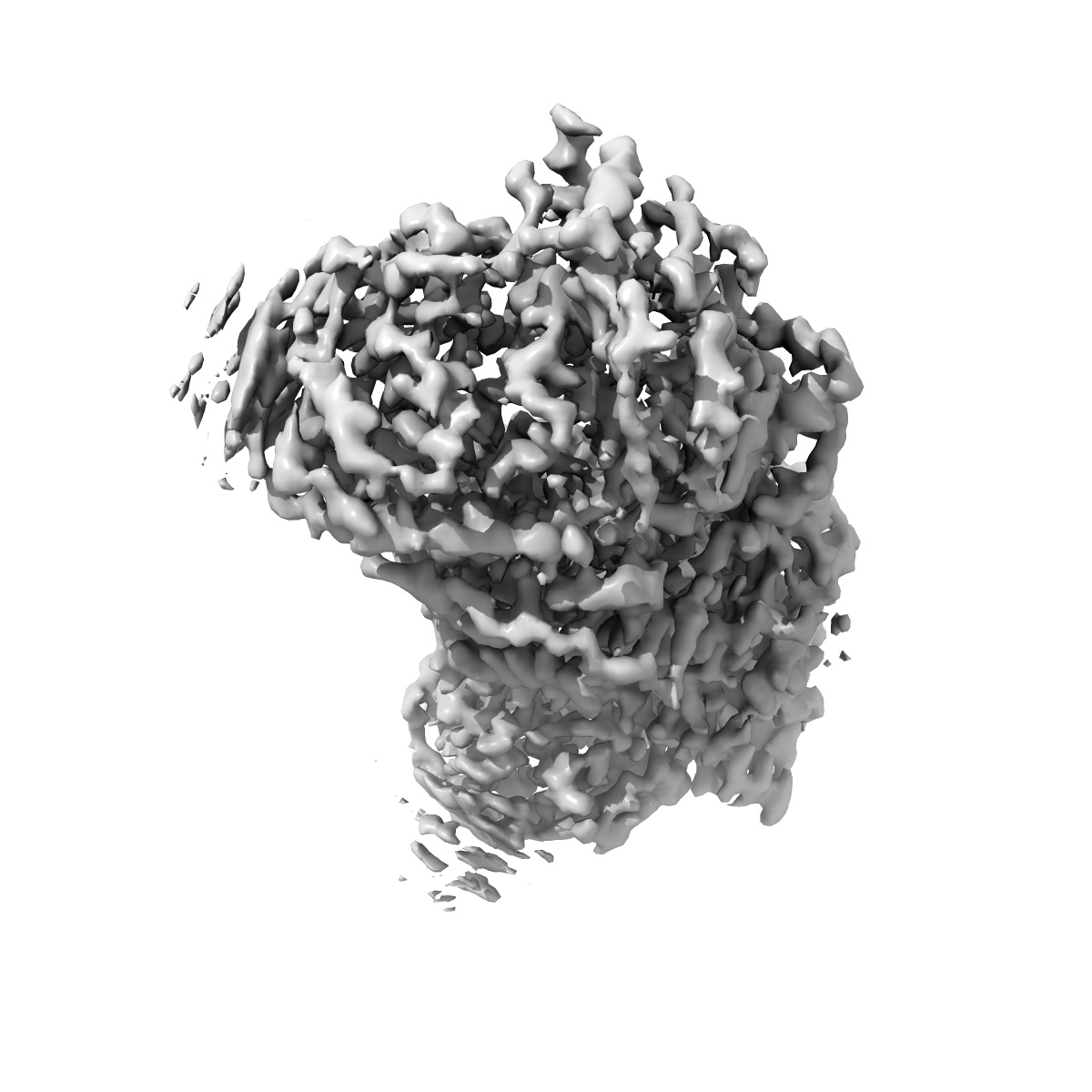
EMD-7041
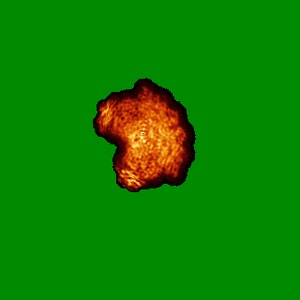
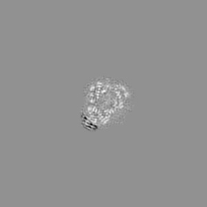
Cryo-EM structure of human insulin degrading enzyme in complex with insulin
EMD-7041
Single-particle3.7 Å
 Deposition: 22/09/2017
Map released: 22/11/2017
Last modified: 13/11/2024
Concentration: 0.3
mg/mL
Details: The sample was monodisperse
Details: The sample was monodisperse
Buffer
pH: 7.8
Buffer components [3]:
Buffer components [3]:
| Name | Formula | Concentration | ChEBI |
|---|---|---|---|
| HEPES | C8H18N2O4S | 20.0 mmol/L | |
| Sodium chloride | NaCl | 300.0 mmol/L | |
| EDTA | C10H16N2O8 | 20.0 mmol/L |
Grid
Vitrification
Cryogen name: ETHANE
Chamber humidity: 85%
Chamber temperature: 298 K
Instrument: HOMEMADE PLUNGER
Details: The cryo grids were made using Spotiton and homemade plunger.
Chamber humidity: 85%
Chamber temperature: 298 K
Instrument: HOMEMADE PLUNGER
Details: The cryo grids were made using Spotiton and homemade plunger.
Microscope: FEI TITAN KRIOS
Illumination mode: FLOOD BEAM
Imaging mode: BRIGHT FIELD
Electron source: FIELD EMISSION GUN
Acceleration voltage: 300 kV
C2 aperture diameter: 70.0 µm
Nominal CS: 2.7 mm
Nominal defocus: 0.9400000000000001 µm - 2.2 µm
Nominal magnification: 22500.0
Calibrated magnification: 46598.0
Specimen holder model: FEI TITAN KRIOS AUTOGRID HOLDER
Cooling holder cryogen: NITROGEN
Alignment procedure: COMA FREE ( Residual tilt: 10.0 mrad)
Temperature
Minimum: 70.0
K
Maximum: 70.0 K
Maximum: 70.0 K
Image Recording
[1]
Detector model:
GATAN K2 SUMMIT (4k x 4k)
Detector mode: COUNTING
Dimensions: 3710 pixel x 3838 pixel
Frames per image: 1-50
Number of grids: 3
Number of real images: 3085
Average exposure time: 10.0 s
Average electron dose per image: 71.4 e/Å2
Detector mode: COUNTING
Dimensions: 3710 pixel x 3838 pixel
Frames per image: 1-50
Number of grids: 3
Number of real images: 3085
Average exposure time: 10.0 s
Average electron dose per image: 71.4 e/Å2
Final
reconstruction
Resolution: 3.7
Å
(
BY AUTHOR)
Resolution method: FSC 0.143 CUT-OFF
Number of classed used: 1
Number of images used: 116122
Algorithm: FOURIER SPACE
Resolution method: FSC 0.143 CUT-OFF
Number of classed used: 1
Number of images used: 116122
Algorithm: FOURIER SPACE
⌯ Applied Symmetry
Point group:
C1
Software
[1]
| Name | Version | Details |
|---|---|---|
| RELION | 2.1 | - |
⦨ Initial angle
assignment
Type:
PROJECTION MATCHING
Number of reference projections: 2
Number of reference projections: 2
Software
[1]
| Name | Version | Details |
|---|---|---|
| RELION | 2.0 | - |
⦩ Final angle assignment
Type:
PROJECTION MATCHING
Number of reference projections: 2
Number of reference projections: 2
Software
[1]
| Name | Version | Details |
|---|---|---|
| RELION | 2.0 | - |
Particle selection
[1]
| Selected | Ref. model | Method | Software | Details |
|---|---|---|---|---|
| 762283 | - | - | - | - |
Final 3D classification
Number of classes:
8
Avg. number of members per classes: 18549.0
Avg. number of members per classes: 18549.0
Software
[1]
| Name | Version | Details |
|---|---|---|
| RELION | 2.0 | - |
Format: CCP4
Data type: IMAGE STORED AS FLOATING POINT NUMBER (4 BYTES)
Annotation details: This is the sharpened map from the final, Insulin degrading enzyme in complex with insulin
Data type: IMAGE STORED AS FLOATING POINT NUMBER (4 BYTES)
Annotation details: This is the sharpened map from the final, Insulin degrading enzyme in complex with insulin
⬡ Geometry
| X | Y | Z | |
|---|---|---|---|
| Origin | 0 | 0 | 0 |
| Dimensions (px) | 320 | 320 | 320 |
| Dimensions (Å) | 343.36 | 343.36 | 343.36 |
| Voxel size (Å) | 1.073 | 1.073 | 1.073 |
Contour list
| Primary | Level | Source |
|---|---|---|
| True | 0.053 | AUTHOR |