






EMD-6740
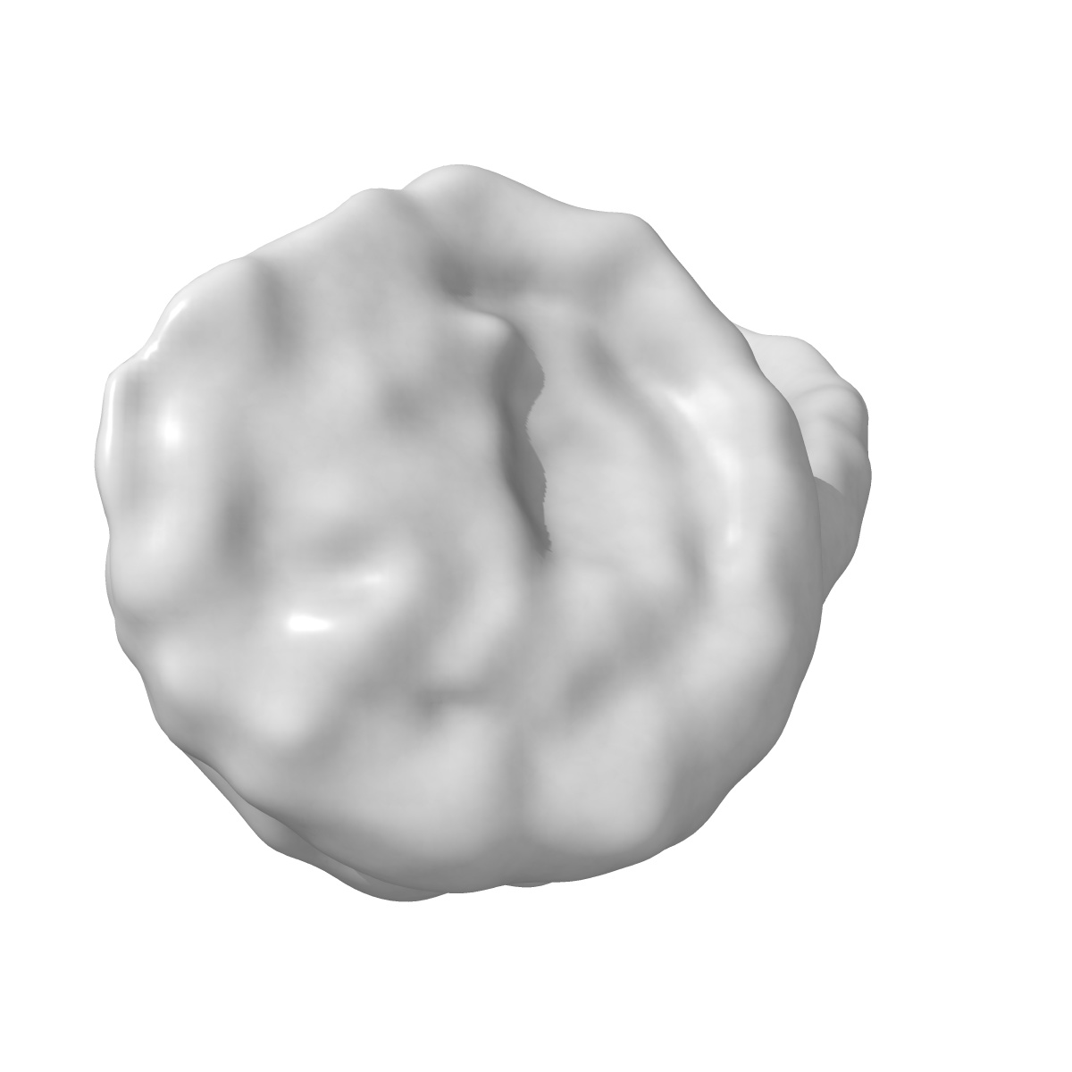
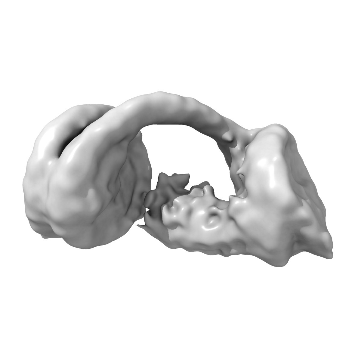
Human HP1alpha-dinucleosome H3K9Me3 class C
EMD-6740
Single-particle11.5 Å
 Deposition: 22/05/2017
Map released: 31/01/2018
Last modified: 05/08/2020
Concentration: .8
mg/mL
Details: Sample was monodisperse
Details: Sample was monodisperse
Buffer
pH: 7.5
Buffer components [2]:
Buffer components [2]:
| Name | Formula | Concentration | ChEBI |
|---|---|---|---|
| Tris-HCl | (HOCH2)3CNH2 | 20.0 mM | |
| DTT | Dithiothreitol | 1.0 mM |
Grid
Vitrification
Cryogen name: ETHANE
Chamber humidity: 100%
Chamber temperature: 289 K
Instrument: FEI VITROBOT MARK IV
Details: 3 second blot, 2.5uL.
Chamber humidity: 100%
Chamber temperature: 289 K
Instrument: FEI VITROBOT MARK IV
Details: 3 second blot, 2.5uL.
Microscope: FEI TITAN KRIOS
Illumination mode: FLOOD BEAM
Imaging mode: BRIGHT FIELD
Electron source: FIELD EMISSION GUN
Acceleration voltage: 300 kV
C2 aperture diameter: 50.0 µm
Nominal CS: 2.62 mm
Nominal magnification: 105000.0
Calibrated magnification: 35971.0
Specimen holder model: FEI TITAN KRIOS AUTOGRID HOLDER
Cooling holder cryogen: NITROGEN
Alignment procedure: COMA FREE ( Residual tilt: 0.1 mrad)
Details: Nanoprobe, parallel beam illumination
Illumination mode: FLOOD BEAM
Imaging mode: BRIGHT FIELD
Electron source: FIELD EMISSION GUN
Acceleration voltage: 300 kV
C2 aperture diameter: 50.0 µm
Nominal CS: 2.62 mm
Nominal magnification: 105000.0
Calibrated magnification: 35971.0
Specimen holder model: FEI TITAN KRIOS AUTOGRID HOLDER
Cooling holder cryogen: NITROGEN
Alignment procedure: COMA FREE ( Residual tilt: 0.1 mrad)
Details: Nanoprobe, parallel beam illumination
Temperature
Minimum: 77.0
K
Maximum: 100.0 K
Maximum: 100.0 K
Specialist optics
Image Recording
[1]
Detector model:
GATAN K2 QUANTUM (4k x 4k)
Detector mode: COUNTING
Dimensions: 3838 pixel x 3710 pixel
Sampling interval: 5.0 µm
Frames per image: 1-40
Number of grids: 2
Number of real images: 2329
Average exposure time: 5.0 s
Average electron dose per image: 38.0 e/Å2
Details: Images were collected with a volta phase plate (VPP) in-focus
Detector mode: COUNTING
Dimensions: 3838 pixel x 3710 pixel
Sampling interval: 5.0 µm
Frames per image: 1-40
Number of grids: 2
Number of real images: 2329
Average exposure time: 5.0 s
Average electron dose per image: 38.0 e/Å2
Details: Images were collected with a volta phase plate (VPP) in-focus
Details: Frame alignment and integration with motioncor2 incl. dose weighting
Final
reconstruction
Resolution: 11.5
Å
(
BY AUTHOR)
Resolution method: FSC 0.143 CUT-OFF
Number of classed used: 1
Number of images used: 21344
Algorithm: FOURIER SPACE
Details:
Resolution method: FSC 0.143 CUT-OFF
Number of classed used: 1
Number of images used: 21344
Algorithm: FOURIER SPACE
Details:
⌯ Applied Symmetry
Point group:
C1
Software
[1]
| Name | Version | Details |
|---|---|---|
| RELION | 2.1 | - |
Startup model
[1]
⦨ Initial angle
assignment
⦩ Final angle assignment
Particle selection
[1]
| Selected | Ref. model | Method | Software | Details |
|---|---|---|---|---|
| 409948 | - | - | - | - |
Final 3D classification
Number of classes:
6
Avg. number of members per classes: 22000.0
Avg. number of members per classes: 22000.0
Software
[1]
| Name | Version | Details |
|---|---|---|
| RELION | 2.1 | - |
CTF correction
Details: In-focus VPP data
Format: CCP4
Data type: IMAGE STORED AS FLOATING POINT NUMBER (4 BYTES)
Data type: IMAGE STORED AS FLOATING POINT NUMBER (4 BYTES)
⬡ Geometry
| X | Y | Z | |
|---|---|---|---|
| Origin | 0 | 0 | 0 |
| Dimensions (px) | 256 | 256 | 256 |
| Dimensions (Å) | 355.84 | 355.84 | 355.84 |
| Voxel size (Å) | 1.39 | 1.39 | 1.39 |
Contour list
| Primary | Level | Source |
|---|---|---|
| True | 3.11 | AUTHOR |