EMD-6479

Single-particle
3.5 Å
EMD-6479 Deposition: 11/10/2015
Map released: 20/01/2016
Last modified: 11/05/2016
Overview 3D View Sample Experiment Validation Additional data Links

EMD-6479

Archive Files (Depositor)

Primary 3D volume (map.gz) Experimental metadata (xml) Experimental metadata (cif.gz)

Validation (wwPDB)

Map-only validation report (pdf.gz) Map-3jck summary report (pdf.gz) Map-3jck FULL report (pdf.gz)

EMDB Annotations

EMICSS entry mapping (xml)

EMDB Files

VA raw map (map)

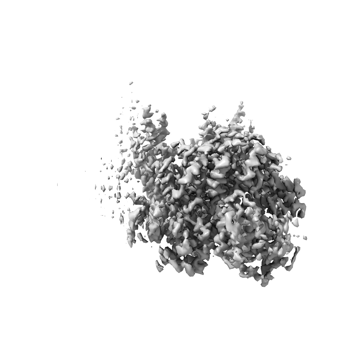
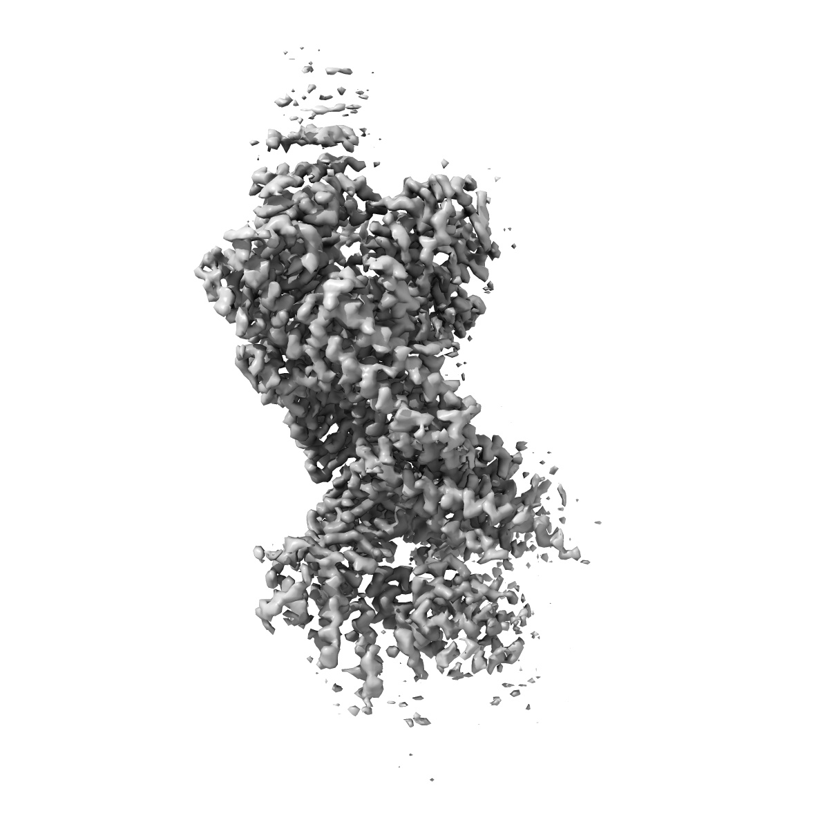
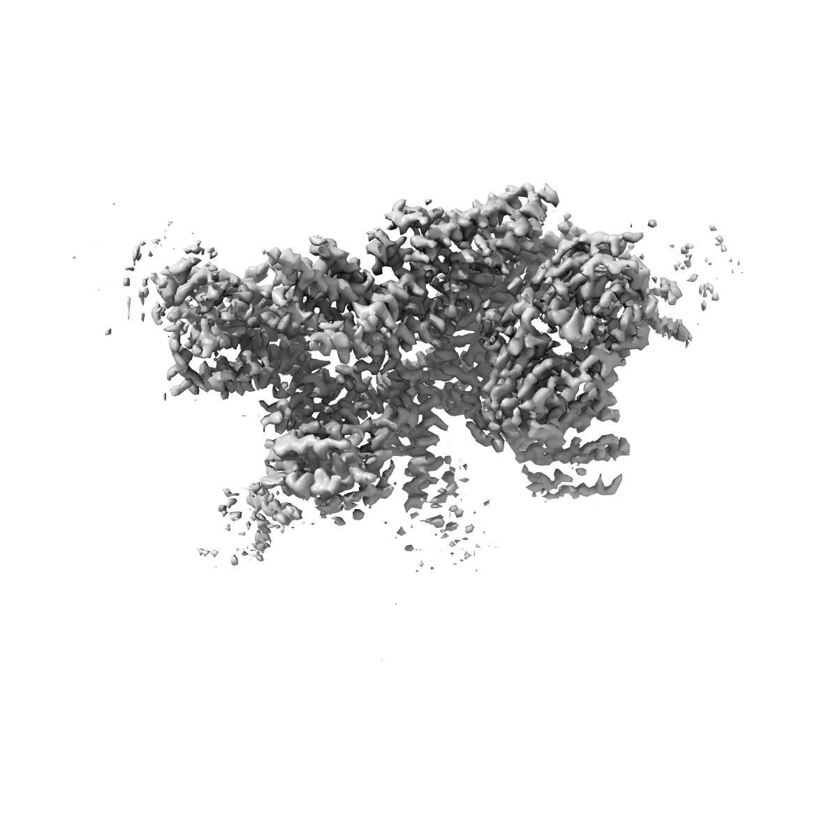
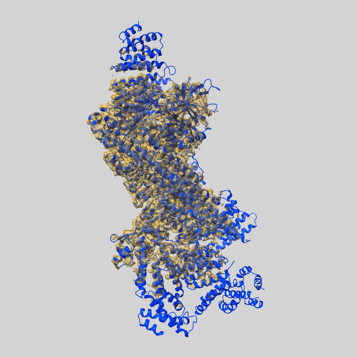
Structure of the yeast 26S proteasome lid sub-complex

EMD-6479

Single-particle
3.5 Å
EMD-6479 Deposition: 11/10/2015
Map released: 20/01/2016
Last modified: 11/05/2016
Overview 3D View Sample Experiment Validation Additional data Links
Sample Organism: Saccharomyces cerevisiae
Sample: Recombinant yeast 26S proteasome lid complex
Fitted models: 3jck

Deposition Authors: Dambacher CM, Worden EJ , Herzik MA , Martin A , Lander GC
Atomic structure of the 26S proteasome lid reveals the mechanism of deubiquitinase inhibition.
Dambacher CM, Worden EJ , Herzik MA , Martin A , Lander GC
(2016) eLife , 5 , e13027 - e13027