EMD-6342

Electron Crystallography
2.5 Å
EMD-6342 Deposition: 29/05/2015
Map released: 10/06/2015
Last modified: 10/06/2015
Overview 3D View Sample Experiment Validation Additional data Links

EMD-6342

Archive Files (Depositor)

Primary 3D volume (map.gz) Half-map 1 Half-map 2 (map.gz) Experimental metadata (xml) Experimental metadata (cif.gz)

Validation (wwPDB)

Map-only validation report (pdf.gz) Map-5a3e summary report (pdf.gz) Map-5a3e FULL report (pdf.gz)

EMDB Annotations

EMICSS entry mapping (xml)

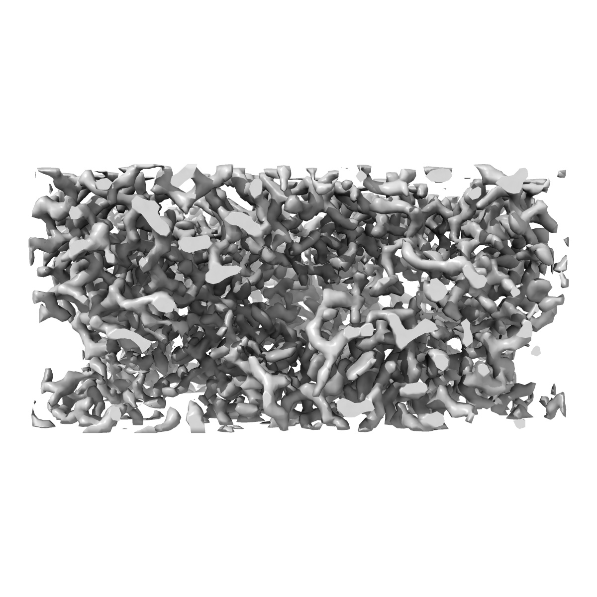
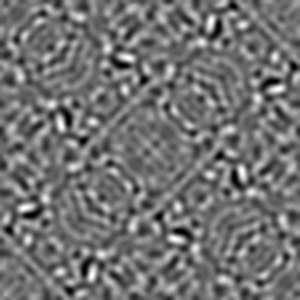
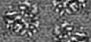
2.5A structure of lysozyme determined by MicroED with data from a single crystal

EMD-6342

Electron Crystallography
2.5 Å
EMD-6342 Deposition: 29/05/2015
Map released: 10/06/2015
Last modified: 10/06/2015
Overview 3D View Sample Experiment Validation Additional data Links
Sample Organism: Gallus gallus
Sample: Hen egg white lysozyme
Fitted models: 5a3e

Deposition Authors: Nannenga BL, Shi D, Leslie AGW, Gonen T
High-resolution structure determination by continuous-rotation data collection in MicroED
Nannenga BL , Shi D, Leslie AG , Gonen T
(2014) Nat. Methods , 11 , 927 - 930