EMD-61003

Single-particle
3.6 Å
EMD-61003 Deposition: 31/07/2024
Map released: 15/01/2025
Last modified: 09/07/2025
Overview 3D View Sample Experiment Validation Additional data Links

EMD-61003

Archive Files (Depositor)

Primary 3D volume (map.gz) Half-map 1 Half-map 2 (map.gz) Experimental metadata (xml) Experimental metadata (cif.gz)

Validation (wwPDB)

Map-only validation report (pdf.gz) Map-9iyx summary report (pdf.gz) Map-9iyx FULL report (pdf.gz)

EMDB Annotations

EMICSS entry mapping (xml)

EMDB Files

VA raw map (map) VA resolution mask (mrc)

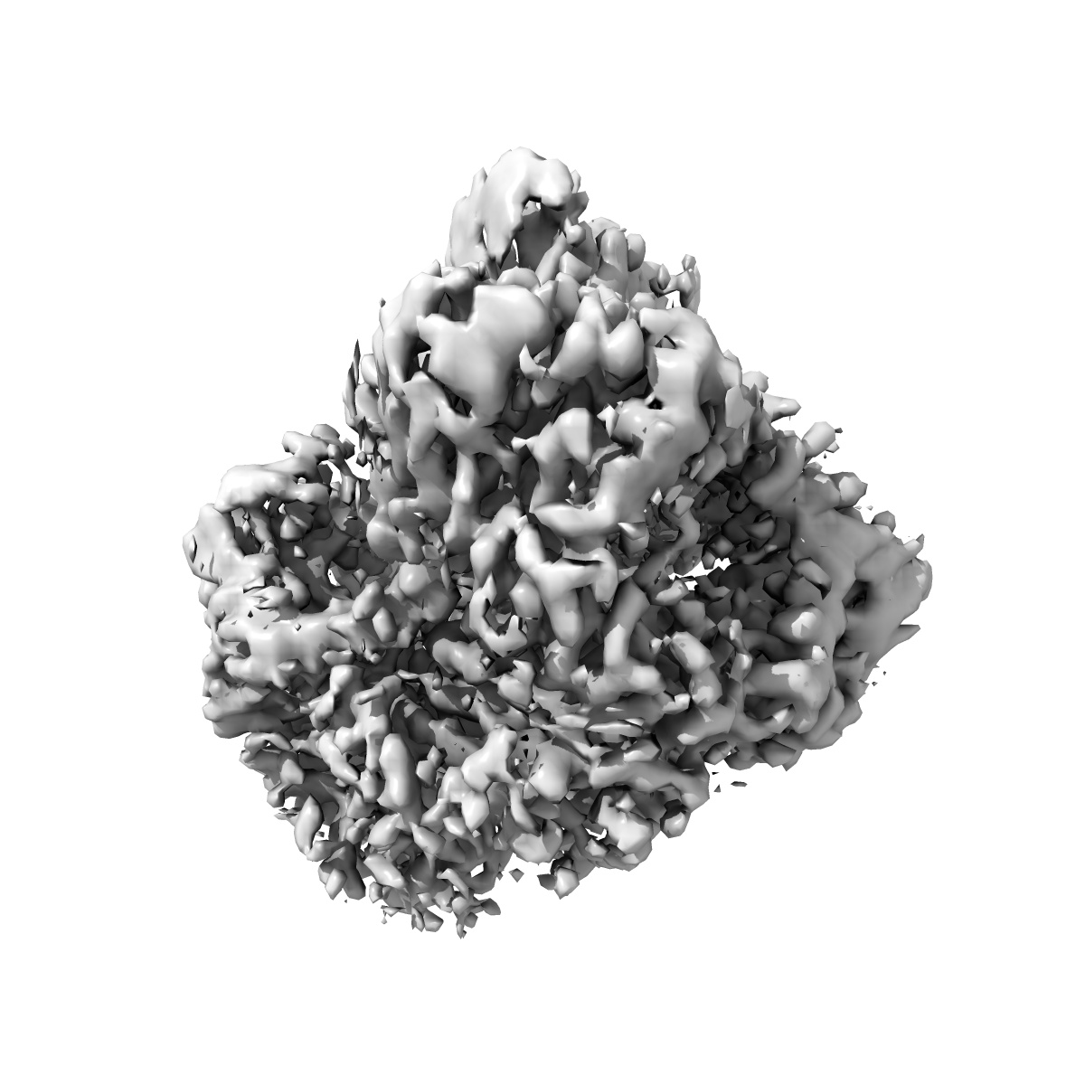
Structure of HBV surface antigen determined in recombinant spherical subviral particle

EMD-61003

Single-particle
3.6 Å
EMD-61003 Deposition: 31/07/2024
Map released: 15/01/2025
Last modified: 09/07/2025
Overview 3D View Sample Experiment Validation Additional data Links
Sample Organism: HBV genotype D3
Sample: HBV surface antigen dimer
Fitted models: 9iyx

Deposition Authors: Chen L , He X
Structure of small HBV surface antigen reveals mechanism of dimer formation.
He X, Kang Y, Tao W, Xu J , Liu X, Chen L
(2025) Cell Discov , 11 , 6 - 6
PUBMED: 39805867
DOI: doi:10.1038/s41421-024-00768-8
ISSN: 2056-5968