EMD-5751

Single-particle
8.0 Å
EMD-5751 Deposition: 26/08/2013
Map released: 18/09/2013
Last modified: 02/03/2016
Overview 3D View Sample Experiment Validation Additional data Links

EMD-5751

Archive Files (Depositor)

Primary 3D volume (map.gz) Experimental metadata (xml) Experimental metadata (cif.gz)

Validation (wwPDB)

Map-only validation report (pdf.gz) Map-3j4k summary report (pdf.gz) Map-3j4k FULL report (pdf.gz)

EMDB Annotations

EMICSS entry mapping (xml)

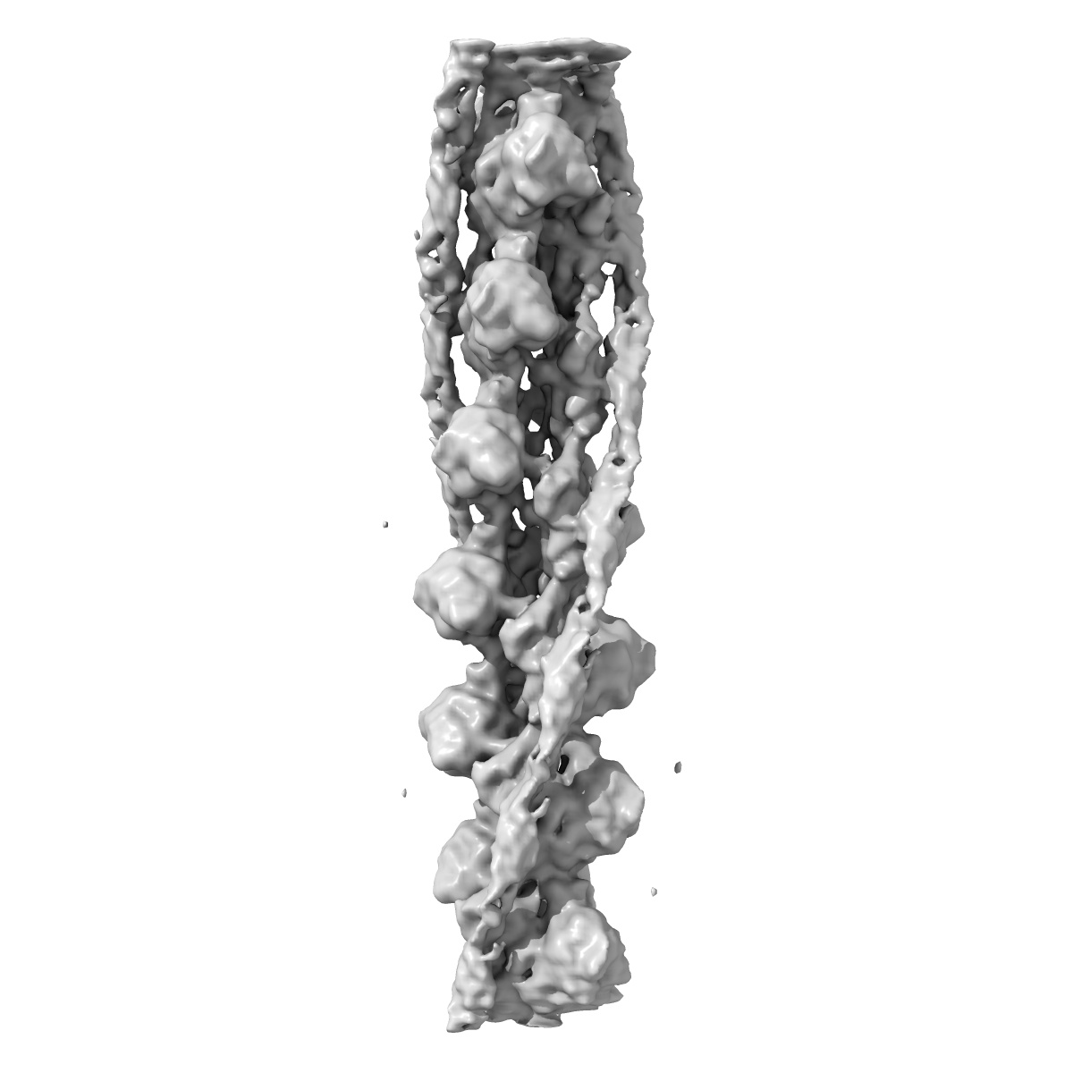
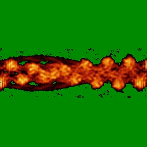
Cryogenic electron microscopy of cardiac actin:tropomyosin

EMD-5751

Single-particle
8.0 Å
EMD-5751 Deposition: 26/08/2013
Map released: 18/09/2013
Last modified: 02/03/2016
Overview 3D View Sample Experiment Validation Additional data Links
Sample Organism: Oryctolagus cuniculus, Gallus gallus
Sample: rabbit skeletal F-actin bound to chicken gizzard tropomyosin
Fitted models: 3j4k

Deposition Authors: Sousa DR, Stagg SM , Stroupe ME
Cryo-EM Structures of the Actin:Tropomyosin Filament Reveal the Mechanism for the Transition from C- to M-State.
Sousa DR, Stagg SM , Stroupe ME
(2013) J. Mol. Biol. , 425 , 4544 - 4555