





EMD-5662
holo-ACP4-PikAIII/C209A/delta ACP5 (no pentaketide)
EMD-5662
Single-particle12.5 Å
 Deposition: 02/05/2013
Map released: 25/06/2014
Last modified: 22/10/2014
Concentration: 0.1
mg/mL
Buffer
pH: 7.4
Details: 50 mM HEPES, 100mM NaCl
Details: 50 mM HEPES, 100mM NaCl
Grid
Details: Glow-discharged Quantifoil R2/200 mesh grid
Vitrification
Cryogen name: ETHANE
Chamber humidity: 100%
Chamber temperature: 89 K
Instrument: FEI VITROBOT MARK IV
Method: Blot for 1.5-2.0 seconds before plunging.
Chamber humidity: 100%
Chamber temperature: 89 K
Instrument: FEI VITROBOT MARK IV
Method: Blot for 1.5-2.0 seconds before plunging.
Microscope: FEI TECNAI F20
Illumination mode: FLOOD BEAM
Imaging mode: BRIGHT FIELD
Electron source: FIELD EMISSION GUN
Acceleration voltage: 120 kV
Nominal CS: 2 mm
Nominal defocus: 1.5 µm - 3.5 µm
Nominal magnification: 50000.0
Calibrated magnification: 66964.0
Specimen holder model: OTHER
Alignment procedure: LEGACY (Astigmatism: Objective lens astigmatism was corrected at 135,000 times magnification, Electron beam tilt params: )
Illumination mode: FLOOD BEAM
Imaging mode: BRIGHT FIELD
Electron source: FIELD EMISSION GUN
Acceleration voltage: 120 kV
Nominal CS: 2 mm
Nominal defocus: 1.5 µm - 3.5 µm
Nominal magnification: 50000.0
Calibrated magnification: 66964.0
Specimen holder model: OTHER
Alignment procedure: LEGACY (Astigmatism: Objective lens astigmatism was corrected at 135,000 times magnification, Electron beam tilt params: )
Temperature
Minimum: 89
K
Maximum: 89 K
Maximum: 89 K
Image Recording
[1]
Detector category:
CCD
Detector model: GATAN ULTRASCAN 4000 (4k x 4k)
Number of real images: 90
Average electron dose per image: 20 e/Å2
Detector model: GATAN ULTRASCAN 4000 (4k x 4k)
Number of real images: 90
Average electron dose per image: 20 e/Å2
Details: The particles were selected manually.
Final
reconstruction
Resolution: 12.5
Å
(
BY AUTHOR)
Resolution method: FSC 0.5 CUT-OFF
Number of images used: 8487
Algorithm: OTHER
Details:
Resolution method: FSC 0.5 CUT-OFF
Number of images used: 8487
Algorithm: OTHER
Details:
⌯ Applied Symmetry
Point group:
C2
Software
[1]
| Name | Version | Details |
|---|---|---|
| EMAN1, EMAN2 | - | - |
CTF correction
Details: Each micrograph
Format: CCP4
Data type: IMAGE STORED AS FLOATING POINT NUMBER (4 BYTES)
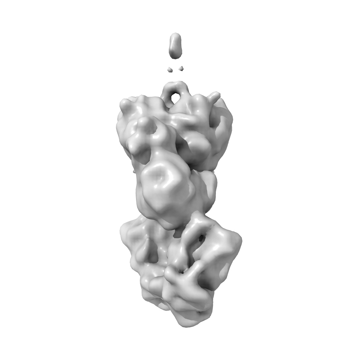
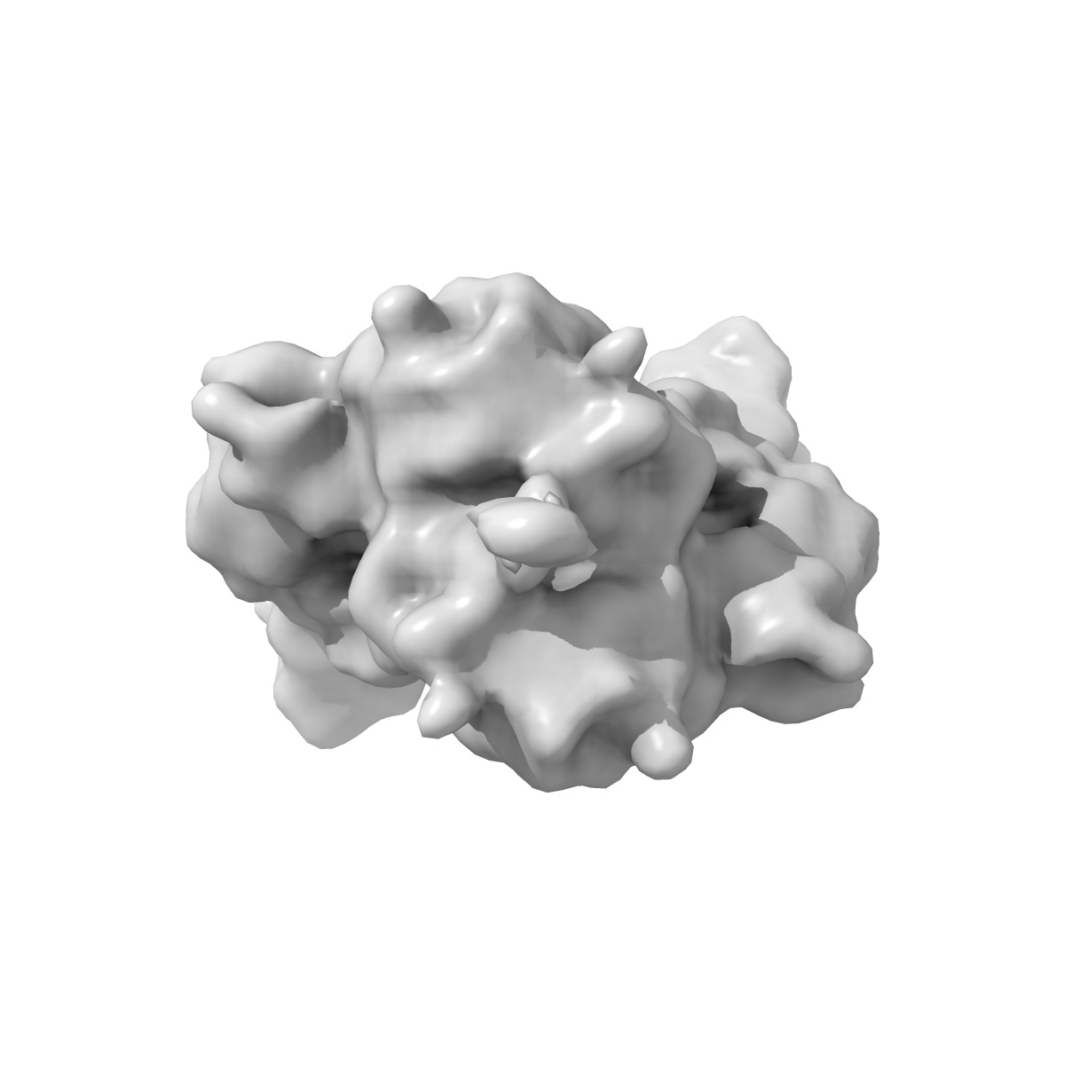
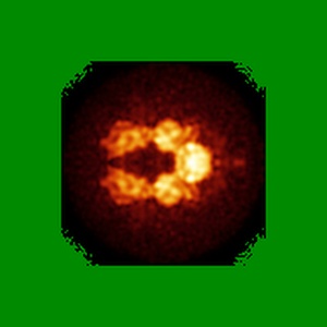
Annotation details: Reconstruction of the 5th module from the pikromycin biosynthetic pathway (PikAIII) lacking the ACP domain with an N-terminal fusion to the ACP domain from module 4 of the pikromycin pathway (ACP4). The ACP4 domain was phosphopantetheinylated.
Details: ::::EMDATABANK.org::::EMD-5662::::
Data type: IMAGE STORED AS FLOATING POINT NUMBER (4 BYTES)
Annotation details: Reconstruction of the 5th module from the pikromycin biosynthetic pathway (PikAIII) lacking the ACP domain with an N-terminal fusion to the ACP domain from module 4 of the pikromycin pathway (ACP4). The ACP4 domain was phosphopantetheinylated.
Details: ::::EMDATABANK.org::::EMD-5662::::
⬡ Geometry
| X | Y | Z | |
|---|---|---|---|
| Origin | -36 | -36 | -36 |
| Dimensions (px) | 192 | 192 | 192 |
| Dimensions (Å) | 430.08002 | 430.08002 | 430.08002 |
| Voxel size (Å) | 2.24 | 2.24 | 2.24 |
Contour list
| Primary | Level | Source |
|---|---|---|
| True | 6.2 | AUTHOR |