
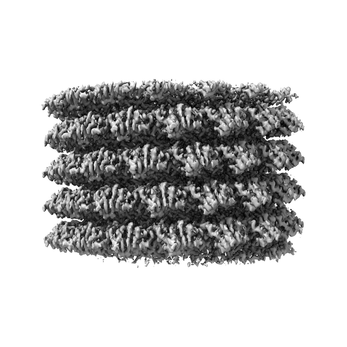

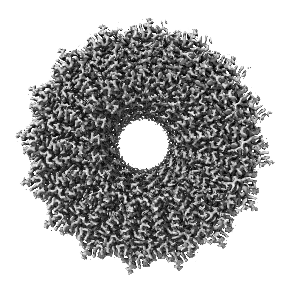





EMD-4413
Tobacco Mosaic Virus
EMD-4413
Helical reconstruction2.3 Å
 Deposition: 13/11/2018
Map released: 20/02/2019
Last modified: 15/05/2024
Concentration: 10
mg/mL
Buffer
pH: 7.0
Grid
Vitrification
Cryogen name: ETHANE
Chamber humidity: 100%
Chamber temperature: 277 K
Instrument: FEI VITROBOT MARK IV
Details: Quantifoil 400 mesh copper grids R 1.2/1.3 were used (Quantifoil Micro Tools GmbH, Jena, Germany). Grids were glow discharged in air at a pressure of 2.2x10⁻² Torr for 2 min at medium power with a Harrick Plasma cleaner (PDC-002). Subsequently, 3-4 ul of the sample was pipetted onto the glow discharged grids and plunge frozen in liquid Ethane with a Vitrobot IV (FEI). The sample chamber of the Vitrobot was maintained at 4C and 100% humidity. Wait time between sample application and blotting was 45 s, the drain time 0 s, the blot time 3 s and the blot force 0..
Chamber humidity: 100%
Chamber temperature: 277 K
Instrument: FEI VITROBOT MARK IV
Details: Quantifoil 400 mesh copper grids R 1.2/1.3 were used (Quantifoil Micro Tools GmbH, Jena, Germany). Grids were glow discharged in air at a pressure of 2.2x10⁻² Torr for 2 min at medium power with a Harrick Plasma cleaner (PDC-002). Subsequently, 3-4 ul of the sample was pipetted onto the glow discharged grids and plunge frozen in liquid Ethane with a Vitrobot IV (FEI). The sample chamber of the Vitrobot was maintained at 4C and 100% humidity. Wait time between sample application and blotting was 45 s, the drain time 0 s, the blot time 3 s and the blot force 0..
Microscope: FEI TITAN KRIOS
Illumination mode: FLOOD BEAM
Imaging mode: BRIGHT FIELD
Electron source: FIELD EMISSION GUN
Acceleration voltage: 300 kV
C2 aperture diameter: 70.0 µm
Nominal CS: 2.7 mm
Nominal defocus: 1.0 µm - 1.8 µm
Calibrated defocus: 0.58 µm - 2.12 µm
Nominal magnification: 75000.0
Specimen holder model: FEI TITAN KRIOS AUTOGRID HOLDER
Cooling holder cryogen: NITROGEN
Alignment procedure: COMA FREE
Illumination mode: FLOOD BEAM
Imaging mode: BRIGHT FIELD
Electron source: FIELD EMISSION GUN
Acceleration voltage: 300 kV
C2 aperture diameter: 70.0 µm
Nominal CS: 2.7 mm
Nominal defocus: 1.0 µm - 1.8 µm
Calibrated defocus: 0.58 µm - 2.12 µm
Nominal magnification: 75000.0
Specimen holder model: FEI TITAN KRIOS AUTOGRID HOLDER
Cooling holder cryogen: NITROGEN
Alignment procedure: COMA FREE
Temperature
Minimum: 79.0
K
Maximum: 80.0 K
Maximum: 80.0 K
Image Recording
[1]
Detector model:
FEI FALCON III (4k x 4k)
Detector mode: INTEGRATING
Dimensions: 4096 pixel x 4096 pixel
Number of grids: 1
Number of real images: 1001
Average exposure time: 5.0 s
Average electron dose per image: 55.0 e/Å2
Detector mode: INTEGRATING
Dimensions: 4096 pixel x 4096 pixel
Number of grids: 1
Number of real images: 1001
Average exposure time: 5.0 s
Average electron dose per image: 55.0 e/Å2
Details: Movies were motion corrected and exposure weighted either with motioncor2 (see tables 1-3 for details). The defocus was determined for the motion corrected and exposure weighted averages with ctffind4
Final
reconstruction
Resolution: 2.3
Å
(
BY AUTHOR)
Resolution method: FSC 0.143 CUT-OFF
Number of classed used: 1
Number of images used: 91927
Algorithm: FOURIER SPACE
Details:
Resolution method: FSC 0.143 CUT-OFF
Number of classed used: 1
Number of images used: 91927
Algorithm: FOURIER SPACE
Details:
⌯ Applied Symmetry
Software
[1]
| Name | Version | Details |
|---|---|---|
| RELION | 3.0 | - |
Startup model
[1]
⦩ Final angle assignment
Segment selection
[1]
| Number selected | Segment length | Segment overlap | Total filament length | Details |
|---|---|---|---|---|
| 393692 | - | - | - | For TMV, some helices were picked manually with the helix picker implemented in relion 2.1. An initial 3D model of these segments was determined using the relion helix option with a cylinder as the starting reference. The aligned helical segments after 3D refinement were subjected to 2D classification without alignment. Three of the 2D class averages were selected as templates for automatic picking of the TMV segments in relion. |
Format: CCP4
Data type: IMAGE STORED AS FLOATING POINT NUMBER (4 BYTES)
Data type: IMAGE STORED AS FLOATING POINT NUMBER (4 BYTES)
⬡ Geometry
| X | Y | Z | |
|---|---|---|---|
| Origin | 0 | 0 | 0 |
| Dimensions (px) | 600 | 600 | 600 |
| Dimensions (Å) | 638.10004 | 638.10004 | 638.10004 |
| Voxel size (Å) | 1.0635 | 1.0635 | 1.0635 |
Contour list
| Primary | Level | Source |
|---|---|---|
| True | 0.05 | AUTHOR |