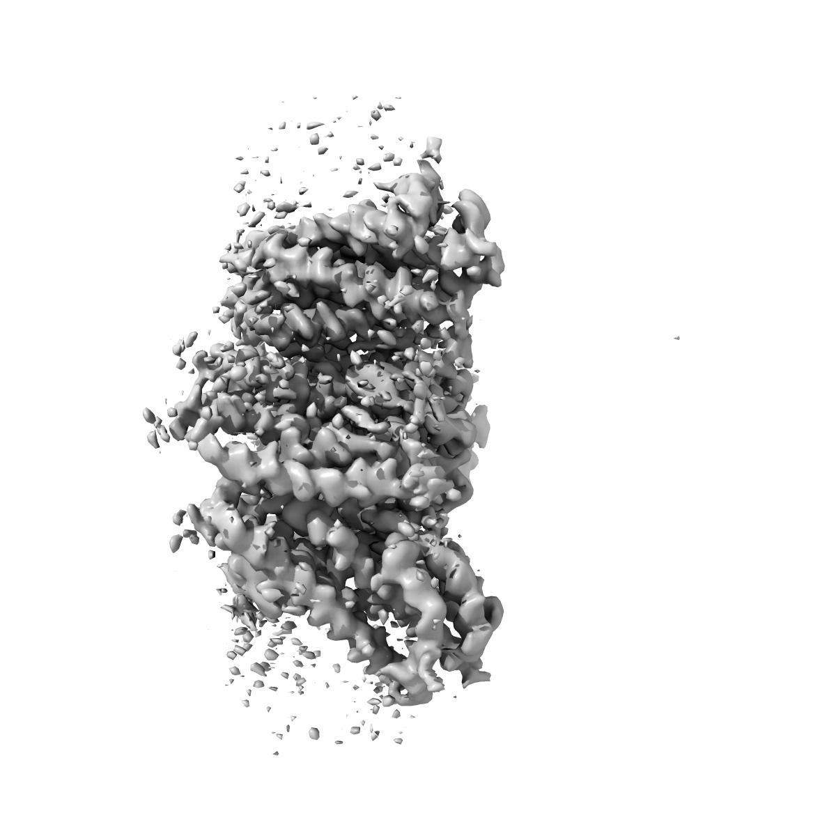
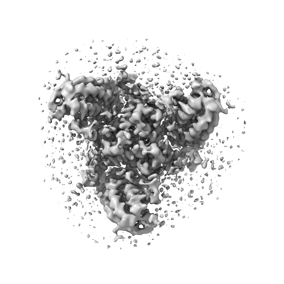
EMD-4386
cryo-EM structure of the human neutral amino acid transporter ASCT2
EMD-4386
Single-particle3.85 Å
 Deposition: 19/04/2018
Map released: 13/06/2018
Last modified: 15/05/2024
Concentration: 2.5
mg/mL
Buffer
pH: 7.0
Details: 20mM Tris-HCl pH 7.4 300mM NaCl 1mM L-glutamine 0.05% DDM 0.005% CHS
Details: 20mM Tris-HCl pH 7.4 300mM NaCl 1mM L-glutamine 0.05% DDM 0.005% CHS
Grid
Vitrification
Cryogen name: ETHANE
Chamber humidity: 100%
Chamber temperature: 278 K
Instrument: FEI VITROBOT MARK II
Chamber humidity: 100%
Chamber temperature: 278 K
Instrument: FEI VITROBOT MARK II
Microscope: FEI TALOS ARCTICA
Illumination mode: FLOOD BEAM
Imaging mode: BRIGHT FIELD
Electron source: FIELD EMISSION GUN
Acceleration voltage: 200 kV
C2 aperture diameter: 100.0 µm
Nominal CS: 2.7 mm
Nominal defocus: 0.0004 µm - 0.0025 µm
Calibrated defocus: 0.0004 µm - 0.0025 µm
Nominal magnification: 49407.0
Calibrated magnification: 49407.0
Specimen holder model: FEI TITAN KRIOS AUTOGRID HOLDER
Cooling holder cryogen: NITROGEN
Alignment procedure: COMA FREE
Temperature
Minimum: 70.0
K
Maximum: 90.0 K
Maximum: 90.0 K
Specialist optics
Energy filter
Image Recording
[1]
Detector model:
GATAN K2 SUMMIT (4k x 4k)
Detector mode: COUNTING
Frames per image: 1-60
Average exposure time: 9.0 s
Average electron dose per image: 0.87 e/Å2
Details: Freshly purified protein was concentrated using Vivaspin concentrating devices with a molecular weight cutoff of 100kDa to 2-2.5 mg ml-1. 2.8 ul were applied on holey-carbon cryo-EM grids (Quantifoil Au R1.2-1.3, 200 and 300 mesh), which were prior glow-discharged at 5 mA for 20 s. Grids were blotted for 3-5 s in a Vitrobot (Mark 3, Thermo Fisher) at 20C temperature and 100% humidity, subsequently plunge-frozen in liquid ethane and stored in liquid nitrogen. Cryo-EM data were collected on a 200 keV Talos Arctica microscope (Thermo Fisher) using a post-column energy filter (Gatan) in zero-loss mode, using a 20 eV slit, a 100 um objective aperture, in an automated fashion using EPU software (Thermo Fisher) on a K2 summit detector (Gatan) in counting mode. Cryo-EM images were acquired at a pixel size of 1.012A (calibrated magnification of 49,407x), a defocus range from -0.4 to 2.5 um, an exposure time of 9 sec and a sub-frame exposure time of 150 ms (60 frames), and a total electron dose on the specimen level of about 52 electrons per A2. Best regions on the grid were screened with a self-written script to calculate the ice thickness and data quality was monitored on the fly using the software FOCUS
Final
reconstruction
Resolution: 3.85
Å
(
BY AUTHOR)
Resolution method: FSC 0.143 CUT-OFF
Number of images used: 184080
Resolution method: FSC 0.143 CUT-OFF
Number of images used: 184080
⌯ Applied Symmetry
Point group:
C3
Software
[1]
| Name | Version | Details |
|---|---|---|
| RELION | 2.1 | - |
⦨ Initial angle
assignment
⦩ Final angle assignment
Format: CCP4
Data type: IMAGE STORED AS FLOATING POINT NUMBER (4 BYTES)
Data type: IMAGE STORED AS FLOATING POINT NUMBER (4 BYTES)
⬡ Geometry
| X | Y | Z | |
|---|---|---|---|
| Origin | 0 | 0 | 0 |
| Dimensions (px) | 240 | 240 | 240 |
| Dimensions (Å) | 242.87999 | 242.87999 | 242.87999 |
| Voxel size (Å) | 1.012 | 1.012 | 1.012 |
Contour list
| Primary | Level | Source |
|---|---|---|
| True | 0.036 | AUTHOR |