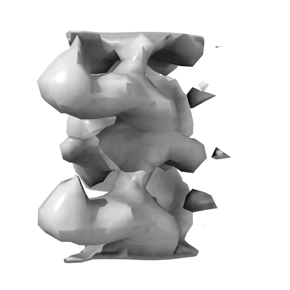
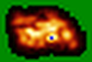
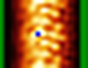
EMD-4304
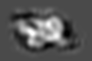
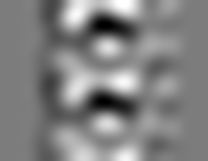
Cryo-EM subtomogram average of IFT complex A in anterograde IFT trains (Chlamydomonas reinhardtii)
EMD-4304
Subtomogram averaging33.0 Å
 Deposition: 21/02/2018
Map released: 31/10/2018
Last modified: 07/11/2018
Microscope: FEI TITAN
Illumination mode: FLOOD BEAM
Imaging mode: BRIGHT FIELD
Electron source: FIELD EMISSION GUN
Acceleration voltage: 300 kV
Illumination mode: FLOOD BEAM
Imaging mode: BRIGHT FIELD
Electron source: FIELD EMISSION GUN
Acceleration voltage: 300 kV
Specialist optics
Energy filter
Name:
GIF Quantum LS
Image Recording
[1]
Detector model:
GATAN K2 SUMMIT (4k x 4k)
Detector mode: SUPER-RESOLUTION
Average electron dose per image: 2.1 e/Å2
Detector mode: SUPER-RESOLUTION
Average electron dose per image: 2.1 e/Å2
Final
reconstruction
Resolution: 33.0
Å
(
BY AUTHOR)
Resolution method: FSC 0.5 CUT-OFF
Resolution method: FSC 0.5 CUT-OFF
Software
[1]
| Name | Version | Details |
|---|---|---|
| PEET | 1.11.0 | - |
⦩ Final angle assignment
Type:
NOT APPLICABLE
Extraction
Number of images used: 750
CTF correction
Software
[1]
| Name | Version | Details |
|---|---|---|
| CTFPHASEFLIP | - | - |
Format: CCP4
Data type: IMAGE STORED AS FLOATING POINT NUMBER (4 BYTES)
Annotation details: Cryo-EM subtomogram average of IFT complex A in anterograde IFT trains (Chlamydomonas reinhartdii)
Data type: IMAGE STORED AS FLOATING POINT NUMBER (4 BYTES)
Annotation details: Cryo-EM subtomogram average of IFT complex A in anterograde IFT trains (Chlamydomonas reinhartdii)
⬡ Geometry
| X | Y | Z | |
|---|---|---|---|
| Origin | -8 | -10 | -7 |
| Dimensions (px) | 22 | 17 | 33 |
| Dimensions (Å) | 310.86002 | 240.21 | 466.29 |
| Voxel size (Å) | 14.130001 | 14.13 | 14.13 |
Contour list
| Primary | Level | Source |
|---|---|---|
| True | 0.512 | AUTHOR |