EMD-4117
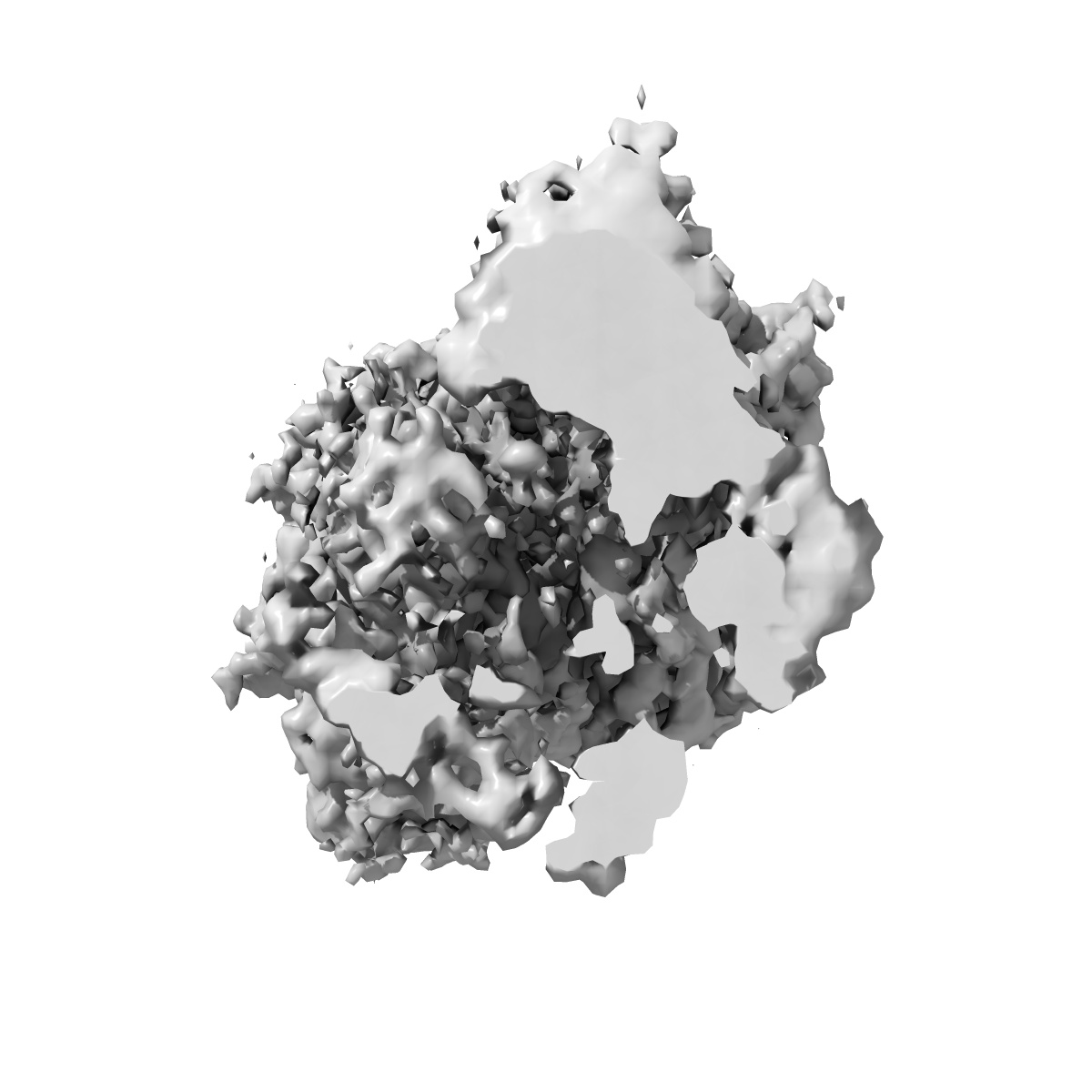
cryoEM structure of crenactin double helical filament at 3.8A resolution
EMD-4117
Helical reconstruction3.8 Å
 Deposition: 23/09/2016
Map released: 18/01/2017
Last modified: 18/01/2017
Buffer
pH: 7.0
Vitrification
Cryogen name: ETHANE
Microscope: FEI POLARA 300
Illumination mode: FLOOD BEAM
Imaging mode: BRIGHT FIELD
Electron source: FIELD EMISSION GUN
Acceleration voltage: 300 kV
Illumination mode: FLOOD BEAM
Imaging mode: BRIGHT FIELD
Electron source: FIELD EMISSION GUN
Acceleration voltage: 300 kV
Image Recording
[1]
Detector model:
FEI FALCON II (4k x 4k)
Detector mode: INTEGRATING
Number of real images: 1474
Average electron dose per image: 40.0 e/Å2
Detector mode: INTEGRATING
Number of real images: 1474
Average electron dose per image: 40.0 e/Å2
Details: Falcon III prototype at 46 frames per second
Final
reconstruction
Resolution: 3.8
Å
(
BY AUTHOR)
Resolution method: FSC 0.143 CUT-OFF
Number of images used: 470396
Details: Helical segments divided at the filament level into the two half sets at the beginning.
Resolution method: FSC 0.143 CUT-OFF
Number of images used: 470396
Details: Helical segments divided at the filament level into the two half sets at the beginning.
⌯ Applied Symmetry
Software
[1]
| Name | Version | Details |
|---|---|---|
| RELION | 2.0 | - |
⦩ Final angle assignment
Type:
NOT APPLICABLE
CTF correction
Software
[1]
| Name | Version | Details |
|---|---|---|
| GCTF | - | - |
Format: CCP4
Data type: IMAGE STORED AS FLOATING POINT NUMBER (4 BYTES)
Data type: IMAGE STORED AS FLOATING POINT NUMBER (4 BYTES)
⬡ Geometry
| X | Y | Z | |
|---|---|---|---|
| Origin | 0 | 0 | 0 |
| Dimensions (px) | 280 | 280 | 280 |
| Dimensions (Å) | 375.2 | 375.2 | 375.2 |
| Voxel size (Å) | 1.34 | 1.34 | 1.34 |
Contour list
| Primary | Level | Source |
|---|---|---|
| True | 0.128 | AUTHOR |