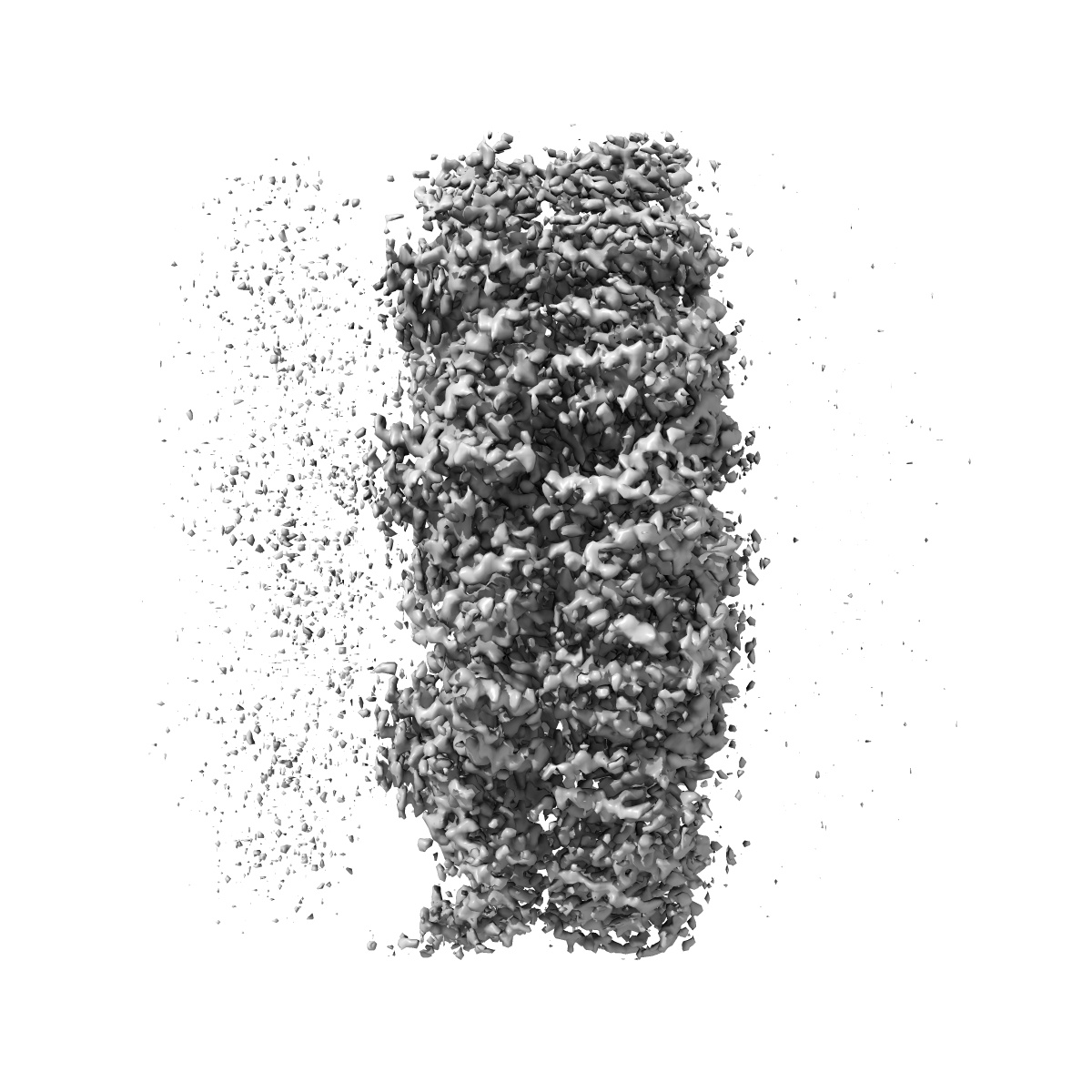
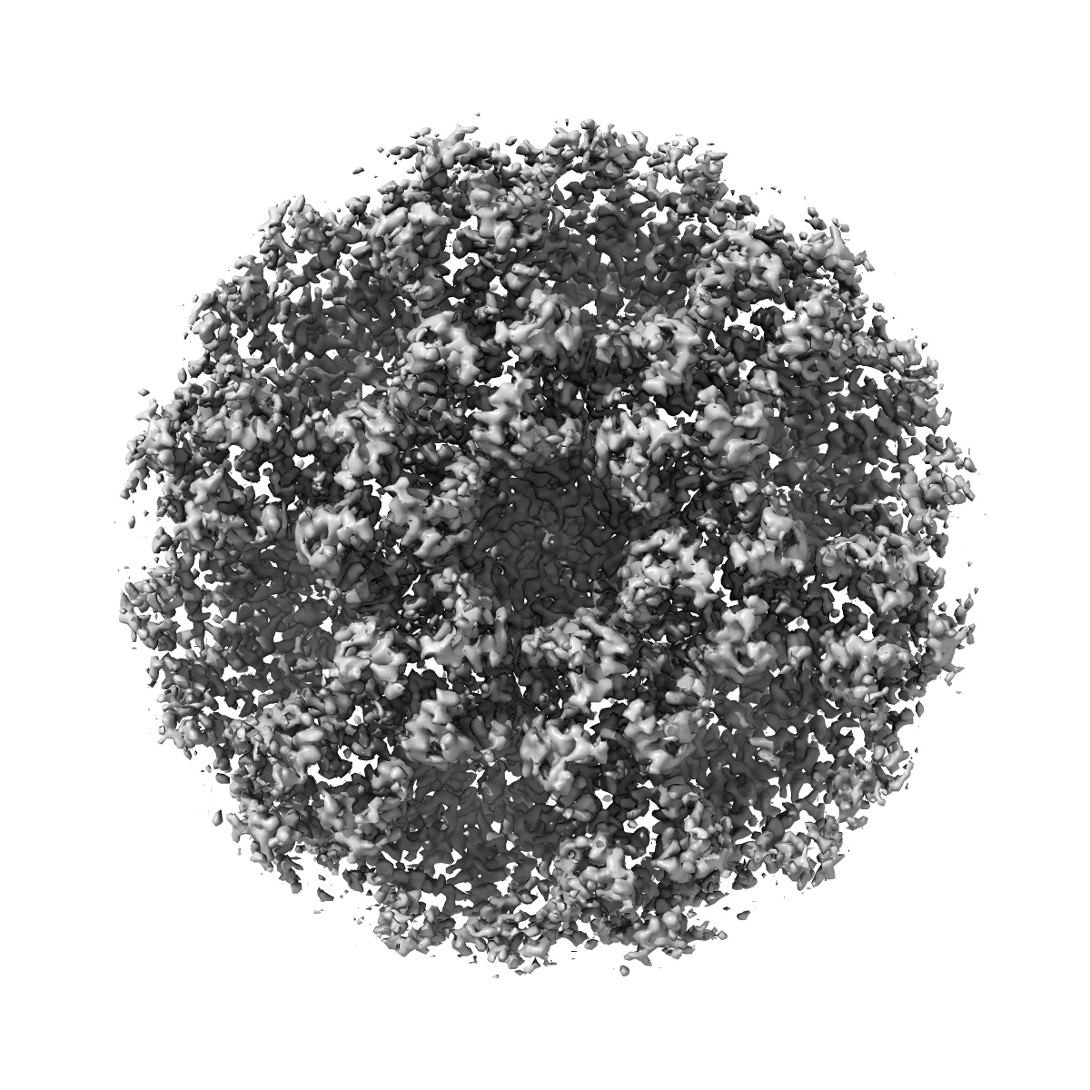
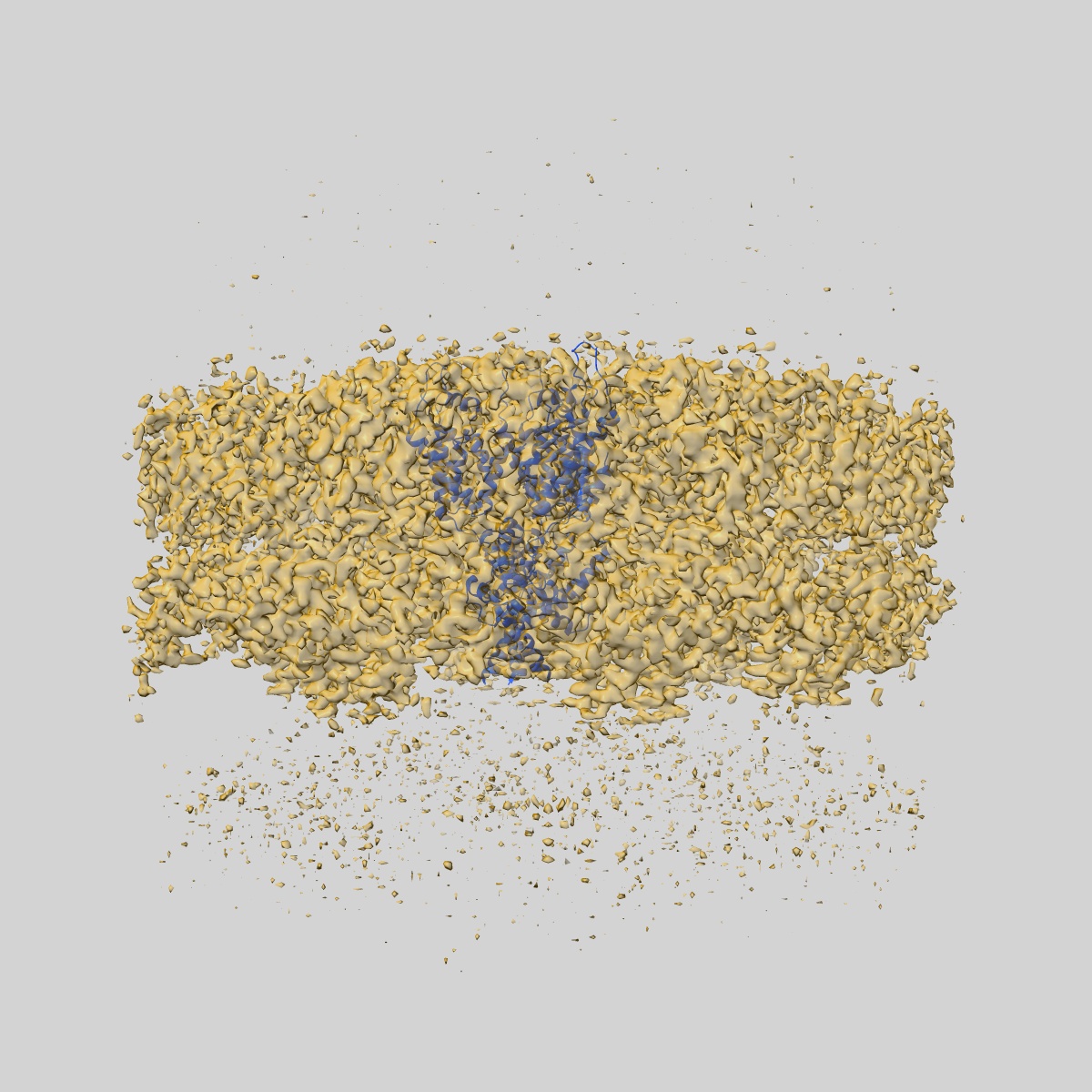
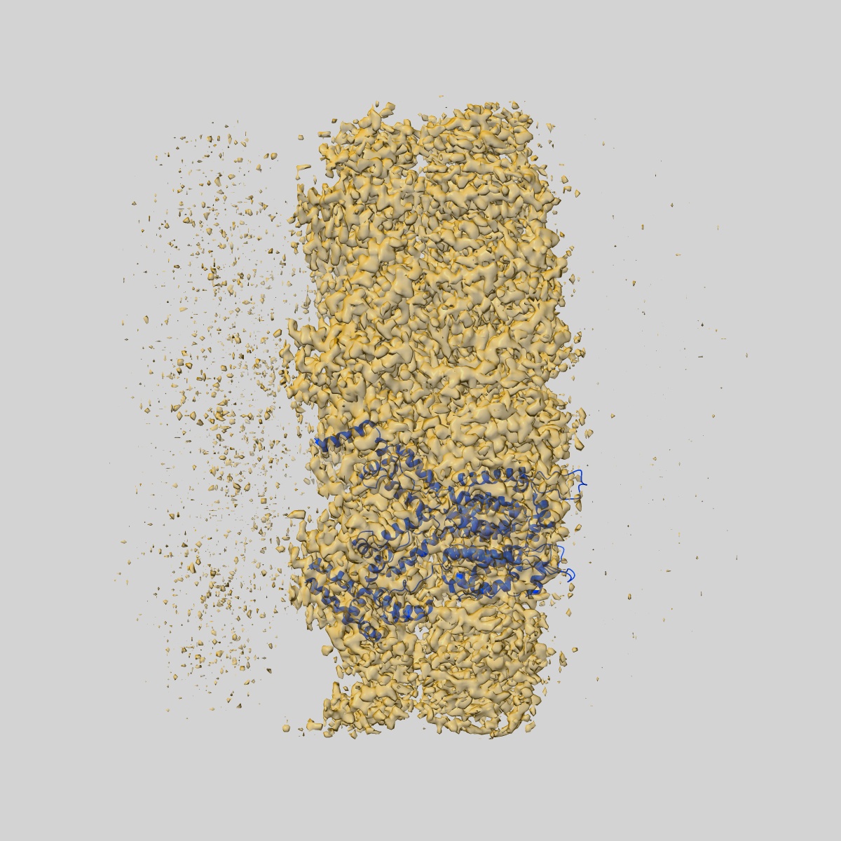
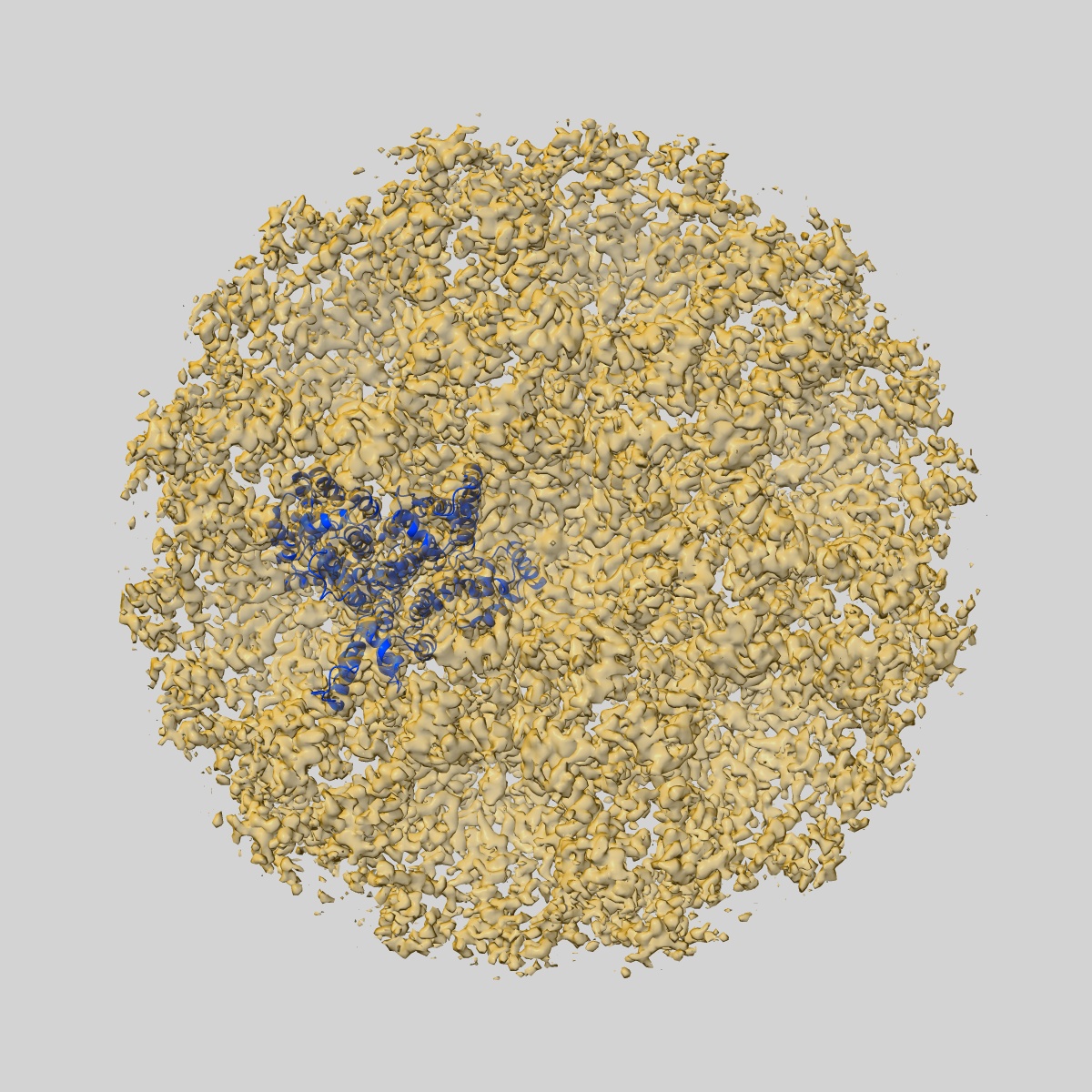
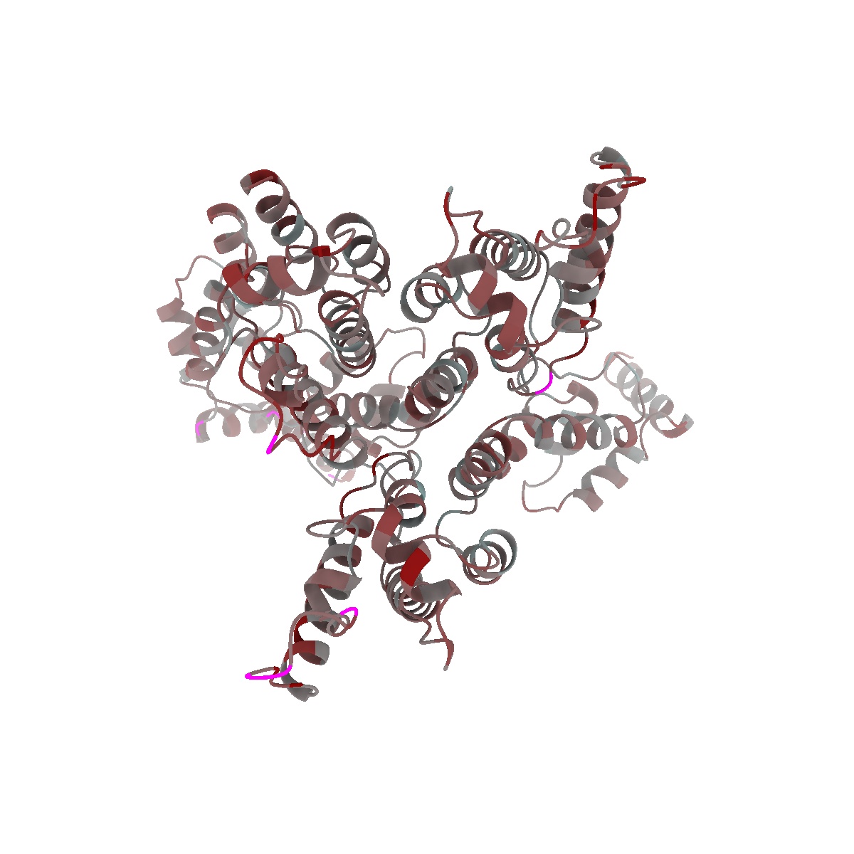
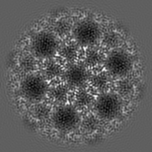
EMD-4015
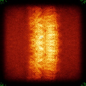
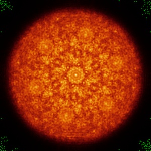
A 3.9 Angstrom structure of HIV-1 CA-SP1 assembled in presence of Bevirimat
EMD-4015
Subtomogram averaging3.9 Å
 Deposition: 09/06/2016
Map released: 13/07/2016
Last modified: 15/05/2024
Concentration: 5
mg/mL
Details: Virus-like particles were assembled in vitro
Details: Virus-like particles were assembled in vitro
Buffer
pH: 8.0
Buffer components [4]:
Details: Virus-like particles were assembled in the presence of nucleic acid (73mer oligonucleotide, 1:10 molar ratio oligonucleotide:protein).
Buffer components [4]:
| Name | Formula | Concentration | ChEBI |
|---|---|---|---|
| HEPES | - | 50.0 mM | - |
| Sodium Chloride | NaCl | 100.0 mM | |
| EDTA | - | 1.0 mM | - |
| TCEP | - | 1.0 mM | - |
Vitrification
Cryogen name: ETHANE
Chamber humidity: 95%
Chamber temperature: 15 K
Instrument: FEI VITROBOT MARK II
Details: 10nM colloidal gold was added to the sample prior to plunge freezing..
Chamber humidity: 95%
Chamber temperature: 15 K
Instrument: FEI VITROBOT MARK II
Details: 10nM colloidal gold was added to the sample prior to plunge freezing..
Microscope: FEI TITAN KRIOS
Illumination mode: FLOOD BEAM
Imaging mode: BRIGHT FIELD
Electron source: FIELD EMISSION GUN
Acceleration voltage: 300 kV
C2 aperture diameter: 50.0 µm
Nominal CS: 2.7 mm
Nominal defocus: 1.5 µm - 5.0 µm
Nominal magnification: 105000.0
Specimen holder model: FEI TITAN KRIOS AUTOGRID HOLDER
Cooling holder cryogen: NITROGEN
Alignment procedure: ZEMLIN TABLEAU
Details: Nanoprobe
Specialist optics
Energy filter
Image Recording
[1]
Detector model:
GATAN K2 QUANTUM (4k x 4k)
Detector mode: SUPER-RESOLUTION
Dimensions: 3710 pixel x 3838 pixel
Frames per image: 8-10
Number of grids: 2
Average exposure time: 1.0 s
Average electron dose per image: 3.4 e/Å2
Details: Number of frames ranged from 8-10 Exposure time per tilt ranged from 0.8 to 1.0 seconds
Detector mode: SUPER-RESOLUTION
Dimensions: 3710 pixel x 3838 pixel
Frames per image: 8-10
Number of grids: 2
Average exposure time: 1.0 s
Average electron dose per image: 3.4 e/Å2
Details: Number of frames ranged from 8-10 Exposure time per tilt ranged from 0.8 to 1.0 seconds
Details: Frames were aligned using MotionCorr. Tilts in a tilt series were exposure filtered for cumulative electron dose. Tomograms were reconstructed using IMOD.
Final
reconstruction
Resolution: 3.9
Å
(
BY AUTHOR)
Resolution method: FSC 0.143 CUT-OFF
Number of classed used: 1
Details:
Resolution method: FSC 0.143 CUT-OFF
Number of classed used: 1
Details:
⌯ Applied Symmetry
Point group:
C6
Software
[2]
| Name | Version | Details |
|---|---|---|
| AV3 | - | - |
| TOM Toolbox | - | - |
⦩ Final angle assignment
Type:
OTHER
Details: Cross-correlation based template matching
Details: Cross-correlation based template matching
Software
[2]
| Name | Version | Details |
|---|---|---|
| AV3 | - | - |
| TOM Toolbox | - | - |
Extraction
Number of images used: 527528
Format: CCP4
Data type: IMAGE STORED AS FLOATING POINT NUMBER (4 BYTES)
Data type: IMAGE STORED AS FLOATING POINT NUMBER (4 BYTES)
⬡ Geometry
| X | Y | Z | |
|---|---|---|---|
| Origin | 0 | 0 | 0 |
| Dimensions (px) | 192 | 192 | 192 |
| Dimensions (Å) | 259.2 | 259.2 | 259.2 |
| Voxel size (Å) | 1.35 | 1.35 | 1.35 |
Contour list
| Primary | Level | Source |
|---|---|---|
| True | 0.112 | AUTHOR |