EMD-3941
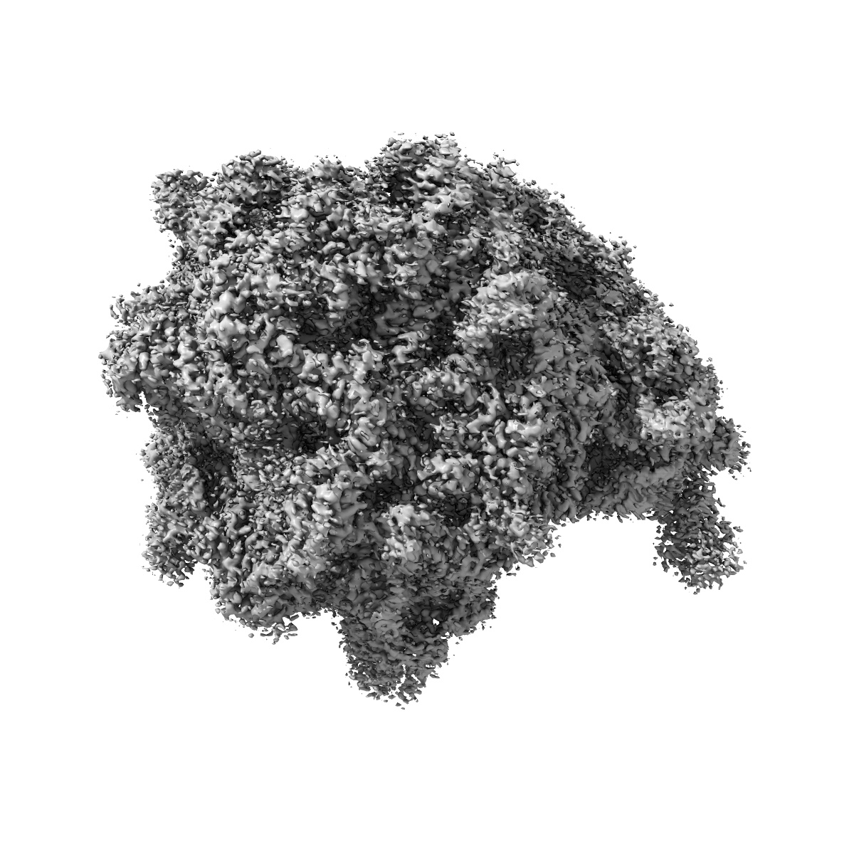
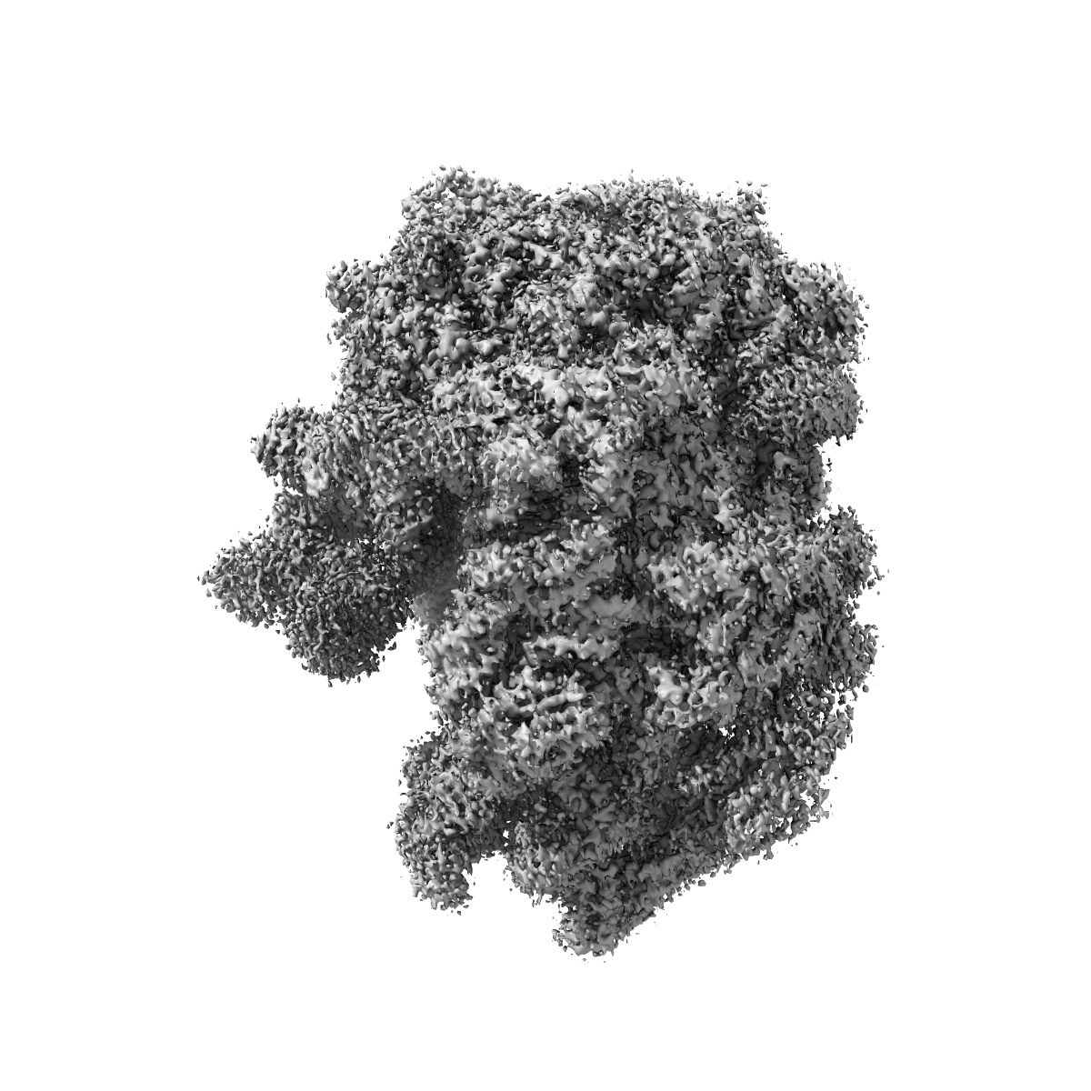
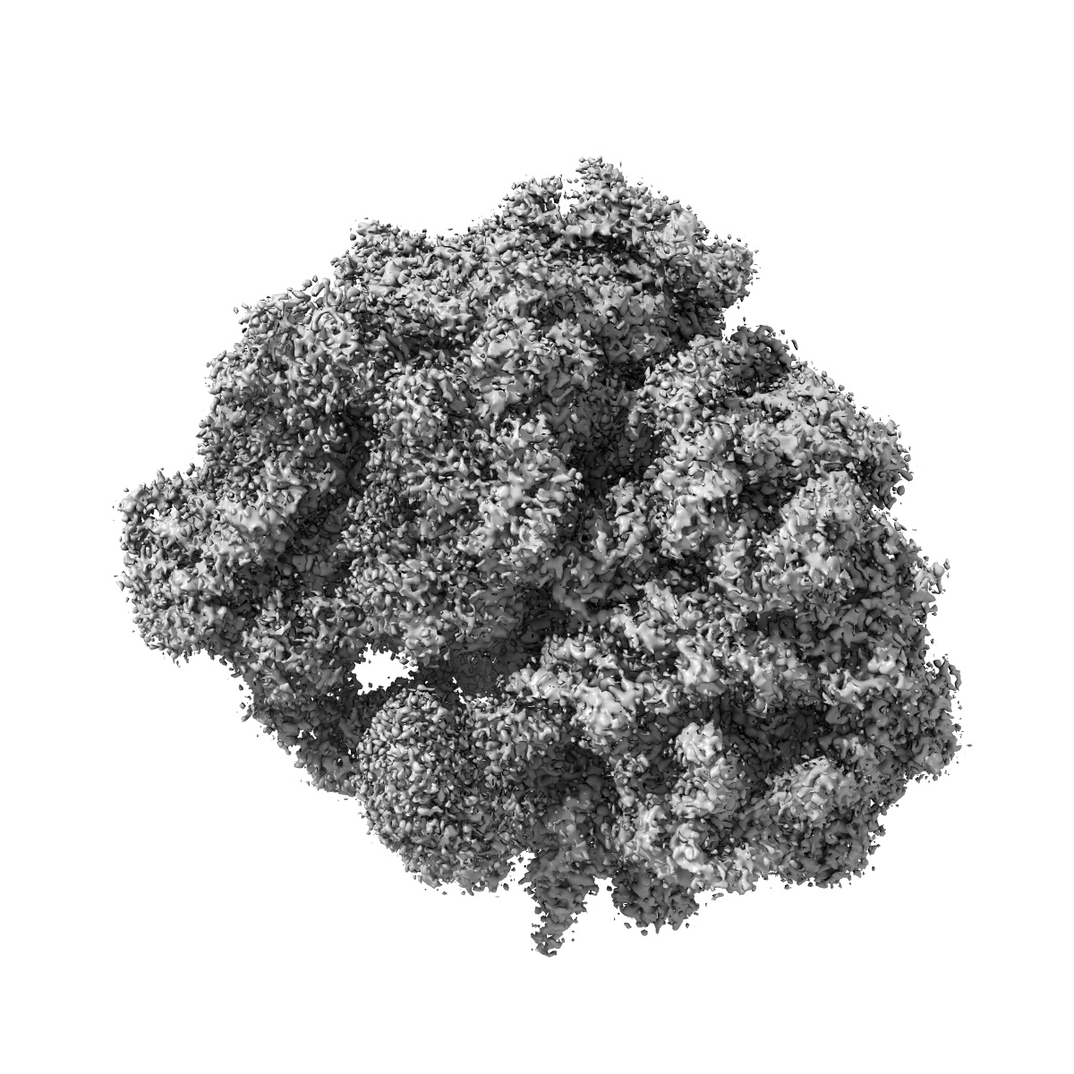
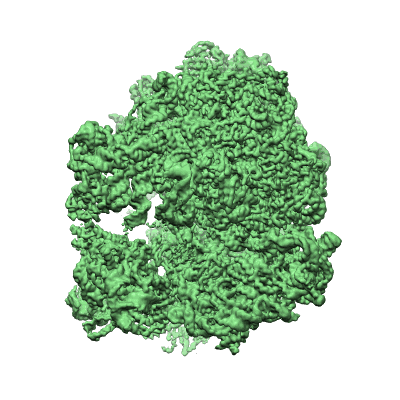
Structure of the chloroplast ribosome with chl-RRF and hibernation-promoting factor
EMD-3941
Single-particle3.0 Å
 Deposition: 18/10/2017
Map released: 18/04/2018
Last modified: 13/11/2024
Buffer
pH: 7.4
Vitrification
Cryogen name: ETHANE
Microscope: FEI TITAN KRIOS
Illumination mode: FLOOD BEAM
Imaging mode: BRIGHT FIELD
Electron source: FIELD EMISSION GUN
Acceleration voltage: 300 kV
Illumination mode: FLOOD BEAM
Imaging mode: BRIGHT FIELD
Electron source: FIELD EMISSION GUN
Acceleration voltage: 300 kV
Image Recording
[1]
Detector model:
GATAN K2 QUANTUM (4k x 4k)
Detector mode: COUNTING
Average electron dose per image: 4.0 e/Å2
Detector mode: COUNTING
Average electron dose per image: 4.0 e/Å2
Final
reconstruction
Resolution: 3.0
Å
(
BY AUTHOR)
Resolution method: FSC 0.143 CUT-OFF
Number of images used: 130300
Resolution method: FSC 0.143 CUT-OFF
Number of images used: 130300
⌯ Applied Symmetry
Point group:
C1
Startup model
[1]
Type:
EMDB MAP
⦨ Initial angle
assignment
Type:
PROJECTION MATCHING
⦩ Final angle assignment
Type:
PROJECTION MATCHING
Format: CCP4
Data type: IMAGE STORED AS FLOATING POINT NUMBER (4 BYTES)
Data type: IMAGE STORED AS FLOATING POINT NUMBER (4 BYTES)
⬡ Geometry
| X | Y | Z | |
|---|---|---|---|
| Origin | 0 | 0 | 0 |
| Dimensions (px) | 420 | 420 | 420 |
| Dimensions (Å) | 445.19998 | 445.19998 | 445.19998 |
| Voxel size (Å) | 1.06 | 1.06 | 1.06 |
Contour list
| Primary | Level | Source |
|---|---|---|
| True | 0.03 | EMDB |