EMD-3809

Helical reconstruction
4.2 Å
EMD-3809 Deposition: 13/07/2017
Map released: 22/11/2017
Last modified: 20/11/2024
Overview 3D View Sample Experiment Validation Additional data Links

EMD-3809

Archive Files (Depositor)

Primary 3D volume (map.gz) Experimental metadata (xml) Experimental metadata (cif.gz)

Validation (wwPDB)

Map-only validation report (pdf.gz) Map-5oh0 summary report (pdf.gz) Map-5oh0 FULL report (pdf.gz)

EMDB Annotations

EMICSS entry mapping (xml)

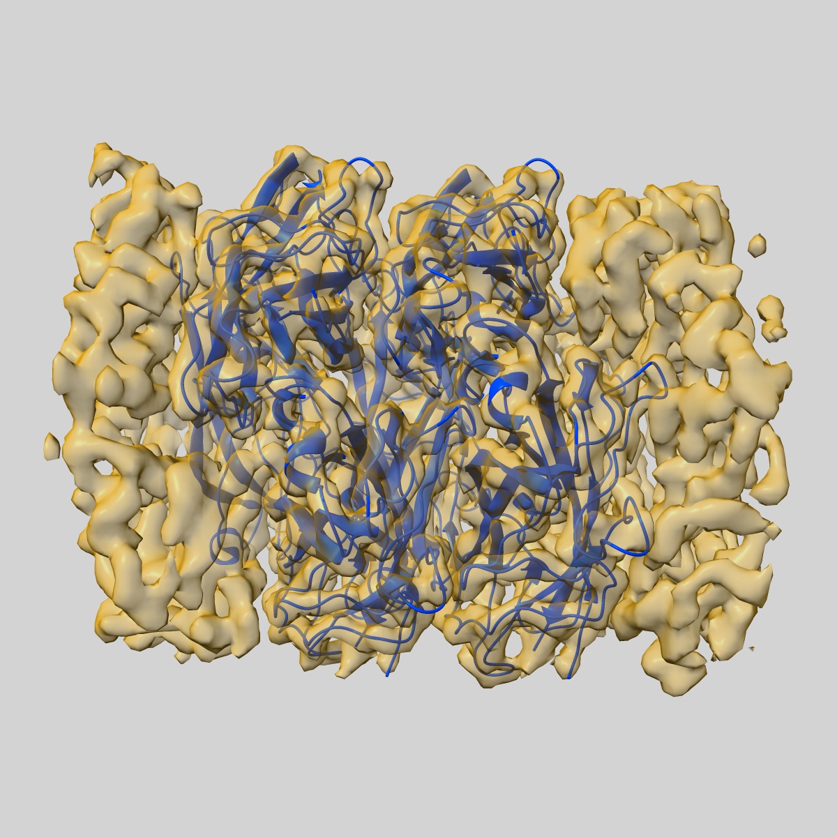
The Cryo-Electron Microscopy Structure of the Type 1 Chaperone-Usher Pilus Rod

EMD-3809

Helical reconstruction
4.2 Å
EMD-3809 Deposition: 13/07/2017
Map released: 22/11/2017
Last modified: 20/11/2024
Overview 3D View Sample Experiment Validation Additional data Links
Sample Organism: Escherichia coli J96
Sample: Type 1 Chaperone-usher pilus
Fitted models: 5oh0

Deposition Authors: Hospenthal MK , Costa TRD , Redzej A , Waksman G
The Cryoelectron Microscopy Structure of the Type 1 Chaperone-Usher Pilus Rod.
Hospenthal MK , Zyla D , Costa TRD , Redzej A , Giese C , Lillington J, Glockshuber R, Waksman G
(2017) Structure , 25 , 1829 - 1838.e4
PUBMED: 29129382
DOI: doi:10.1016/j.str.2017.10.004
ISSN: 1878-4186
ASTM: STRUE6
Grant Support: