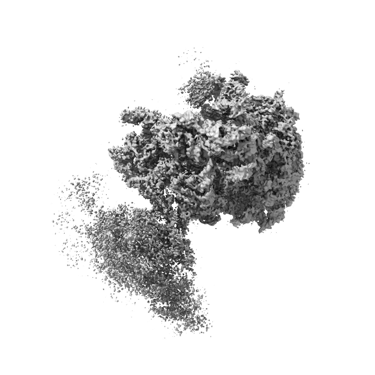
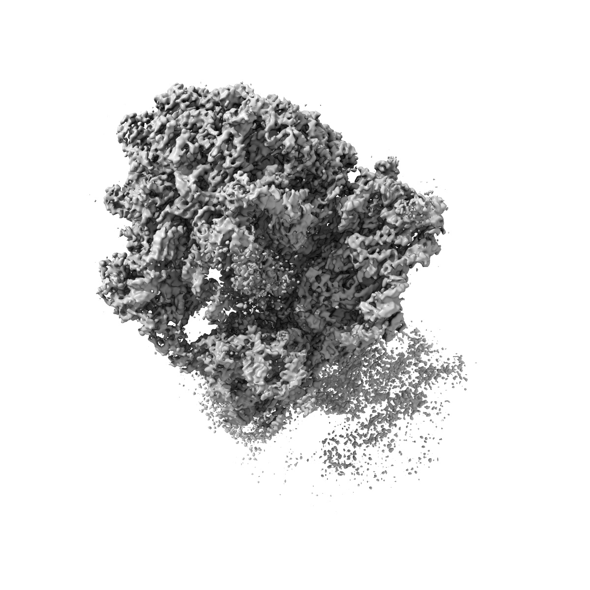
EMD-3656

Single-particle
3.8 Å
EMD-3656 Deposition: 29/03/2017
Map released: 14/06/2017
Last modified: 15/05/2024
Overview 3D View Sample Experiment Validation Additional data Links

EMD-3656

Archive Files (Depositor)

Primary 3D volume (map.gz) Half-map 1 Half-map 2 (map.gz) Experimental metadata (xml) Experimental metadata (cif.gz)

Validation (wwPDB)

Map-only validation report (pdf.gz) Map-5njt summary report (pdf.gz) Map-5njt FULL report (pdf.gz)

EMDB Annotations

EMICSS entry mapping (xml)

EMDB Files

VA raw map (map) VA resolution mask (mrc)

Structure of the Bacillus subtilis hibernating 100S ribosome reveals the basis for 70S dimerization.

EMD-3656

Single-particle
3.8 Å
EMD-3656 Deposition: 29/03/2017
Map released: 14/06/2017
Last modified: 15/05/2024
Overview 3D View Sample Experiment Validation Additional data Links
Sample Organism: Bacillus subtilis subsp. subtilis str. 168
Sample: Structure of the Bacillus subtilis hibernating 100S ribosome reveals the basis for 70S dimerization
Fitted models: 5njt

Deposition Authors: Beckert B, Abdelshahid M, Schaefer H, Steinchen W, Arenz S, Berninghausen O, Beckmann R, Bange G , Turgay K , Wilson DN
Structure of the Bacillus subtilis hibernating 100S ribosome reveals the basis for 70S dimerization.
PUBMED: 28468753
DOI: doi:10.15252/embj.201696189
ISSN: 1460-2075
ASTM: EMJODG
Grant Support: