EMD-3631
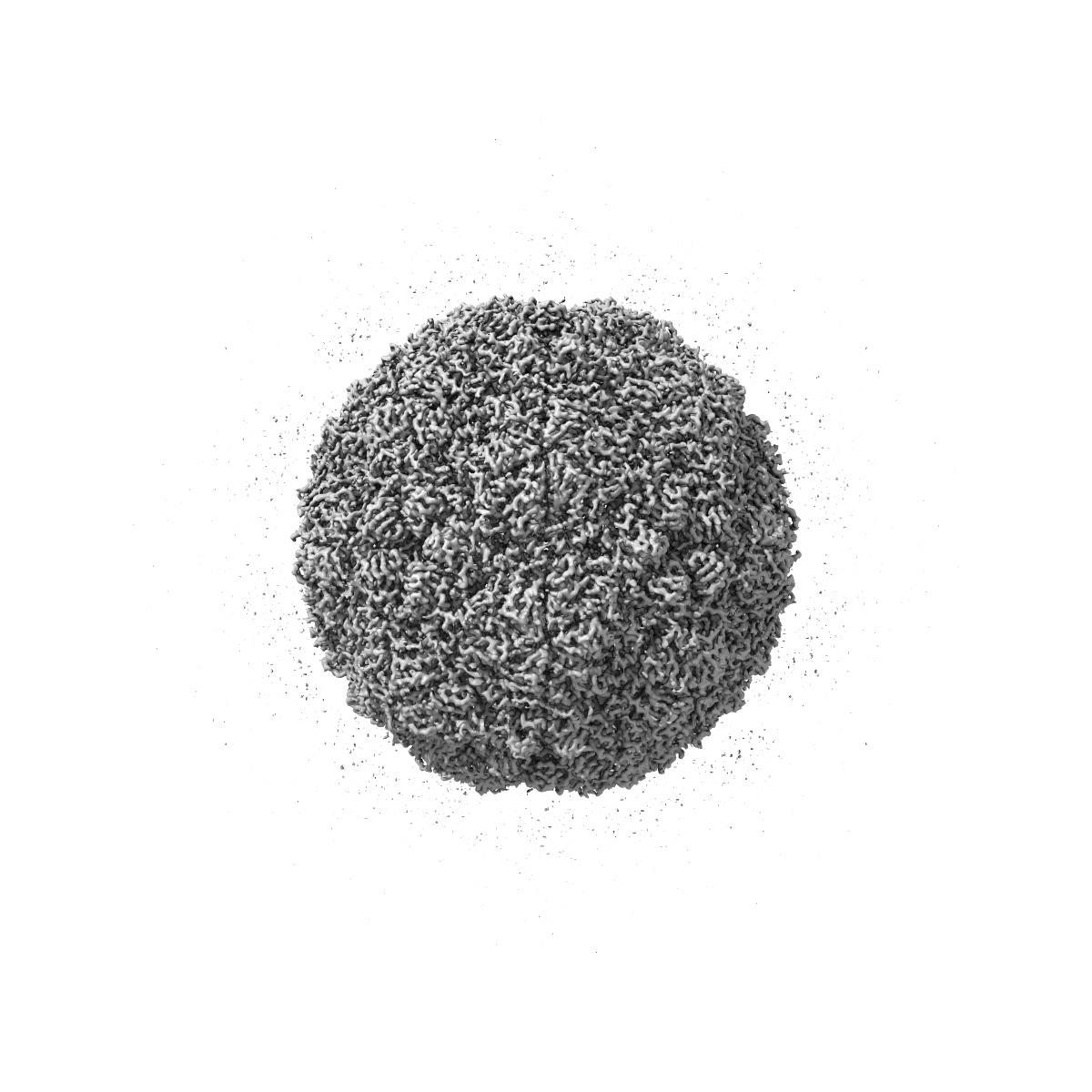
CryoEM Structure of Foot and Mouth Disease Virus O1 Manisa
EMD-3631
Single-particle3.1 Å
 Deposition: 10/03/2017
Map released: 21/06/2017
Last modified: 15/05/2024
Concentration: 0.5
mg/mL
Buffer
pH: 8.0
Buffer components [1]:
Buffer components [1]:
| Name | Formula | Concentration | ChEBI |
|---|---|---|---|
| HEPES | - | 50.0 mM | - |
Grid
Vitrification
Cryogen name: ETHANE
Chamber humidity: 90%
Chamber temperature: 294 K
Instrument: FEI VITROBOT MARK IV
Chamber humidity: 90%
Chamber temperature: 294 K
Instrument: FEI VITROBOT MARK IV
Microscope: FEI POLARA 300
Illumination mode: FLOOD BEAM
Imaging mode: BRIGHT FIELD
Electron source: FIELD EMISSION GUN
Acceleration voltage: 300 kV
C2 aperture diameter: 50.0 µm
Nominal CS: 2.0 mm
Nominal defocus: 1.0 µm - 4.0 µm
Nominal magnification: 160000.0
Calibrated magnification: 37037.0
Specimen holder model: GATAN 910 MULTI-SPECIMEN SINGLE TILT CRYO TRANSFER HOLDER
Cooling holder cryogen: NITROGEN
Alignment procedure: BASIC
Temperature
Minimum: 70.0
K
Maximum: 70.0 K
Maximum: 70.0 K
Specialist optics
Energy filter
Image Recording
[1]
Detector model:
GATAN K2 SUMMIT (4k x 4k)
Detector mode: SUPER-RESOLUTION
Dimensions: 3838 pixel x 3710 pixel
Frames per image: 2-20
Number of grids: 1
Number of real images: 360
Average exposure time: 5.0 s
Average electron dose per image: 18.0 e/Å2
Detector mode: SUPER-RESOLUTION
Dimensions: 3838 pixel x 3710 pixel
Frames per image: 2-20
Number of grids: 1
Number of real images: 360
Average exposure time: 5.0 s
Average electron dose per image: 18.0 e/Å2
Final
reconstruction
Resolution: 3.1
Å
(
BY AUTHOR)
Resolution method: FSC 0.143 CUT-OFF
Number of images used: 13483
Algorithm: BACK PROJECTION
Resolution method: FSC 0.143 CUT-OFF
Number of images used: 13483
Algorithm: BACK PROJECTION
Software
[1]
| Name | Version | Details |
|---|---|---|
| RELION | 1.3 | - |
⦨ Initial angle
assignment
⦩ Final angle assignment
Particle selection
[1]
| Selected | Ref. model | Method | Software | Details |
|---|---|---|---|---|
| 13483 | - | - | - | - |
Final 3D classification
Software
[1]
| Name | Version | Details |
|---|---|---|
| RELION | 1.3 | - |
Format: CCP4
Data type: IMAGE STORED AS FLOATING POINT NUMBER (4 BYTES)
Annotation details: CryoEM Structure of Foot and Mouth Disease Virus O1 Manisa
Data type: IMAGE STORED AS FLOATING POINT NUMBER (4 BYTES)
Annotation details: CryoEM Structure of Foot and Mouth Disease Virus O1 Manisa
⬡ Geometry
| X | Y | Z | |
|---|---|---|---|
| Origin | -199 | -200 | -200 |
| Dimensions (px) | 400 | 400 | 400 |
| Dimensions (Å) | 540.0 | 540.0 | 540.0 |
| Voxel size (Å) | 1.35 | 1.35 | 1.35 |
Contour list
| Primary | Level | Source |
|---|---|---|
| True | 0.037 | AUTHOR |